

Central angiotensin I increases fetal AVP neuron activity and pressor responses
- PMID: 20371731
- PMCID: PMC2886532
- DOI: 10.1152/ajpendo.00060.2010
Central angiotensin I increases fetal AVP neuron activity and pressor responses
Abstract
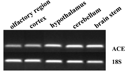
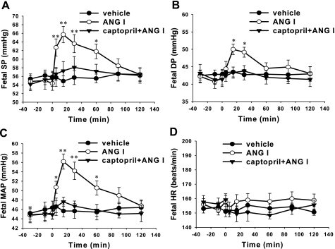
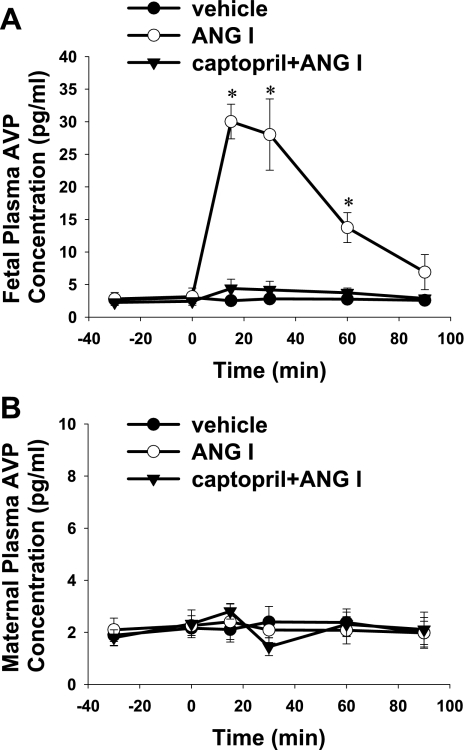
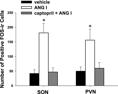
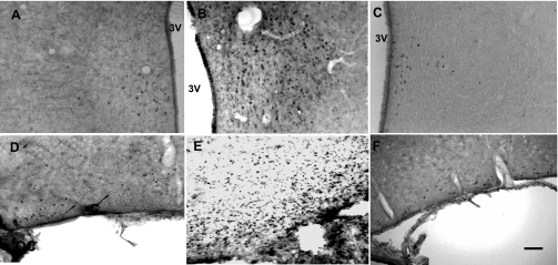
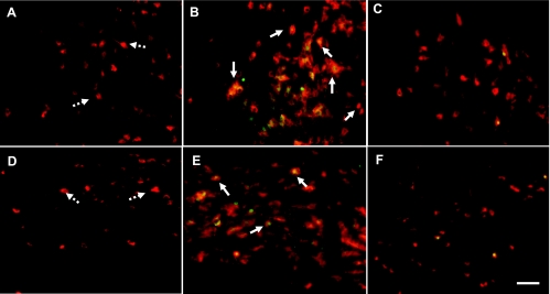
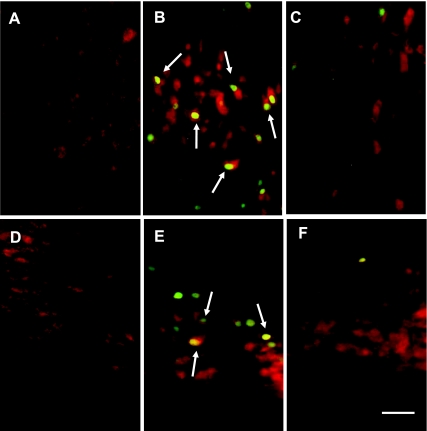
Angiotensin (Ang) II plays a critical role in cardiovascular homeostasis and neuroendocrine regulation. Little is known about whether central angiotensin-converting enzyme (ACE) is functional in the fetal brain. We investigated cardiovascular and neuroendocrinological responses to intracerebroventricular (icv) application of Ang I in the chronically prepared near-term ovine fetus in utero and examined the action sites marked by c-fos expression in the fetal hypothalamus. ACE mRNA was detected in the specific central areas. Intracerebroventricular Ang I significantly increased fetal blood pressure and c-fos expression in the supraoptic nuclei (SON) and the paraventricular nuclei (PVN) in the hypothalamus, accompanied by an increase of fetal plasma arginine vasopressin (AVP). Double labeling demonstrated that AVP neurons in the fetal SON and PVN were expressing c-fos. Captopril, an inhibitor of ACE, significantly suppressed fetal pressor responses and plasma AVP. Double labeling experiments showed colocalization of AT(1) receptor (AT(1)R) and c-fos expression in both SON and PVN following icv Ang I. The results indicate that central endogenous ACE has been functional at least at the last third of gestation and the endogenous brain renin-angiotensin system-mediated pressor responses and AVP release via AT(1)Rs by acting at the sites consistent with the cardiovascular network in the hypothalamus.
Figures
Similar articles
-
In utero development of central ANG-stimulated pressor response and hypothalamic fos expression.Brain Res Dev Brain Res. 2003 Nov 12;145(2):169-76. doi: 10.1016/s0165-3806(03)00226-8. Brain Res Dev Brain Res. 2003. PMID: 14604757
-
Vasopressin mechanism-mediated pressor responses caused by central angiotensin II in the ovine fetus.Pediatr Res. 2004 Nov;56(5):756-62. doi: 10.1203/01.PDR.0000141519.85908.68. Epub 2004 Sep 3. Pediatr Res. 2004. PMID: 15347766
-
Osmotic threshold and sensitivity for vasopressin release and fos expression by hypertonic NaCl in ovine fetus.Am J Physiol Endocrinol Metab. 2000 Dec;279(6):E1207-15. doi: 10.1152/ajpendo.2000.279.6.E1207. Am J Physiol Endocrinol Metab. 2000. PMID: 11093906
-
An overview of the influence of ACE inhibitors on fetal-placental circulation and perinatal development.Mol Cell Biochem. 1997 Nov;176(1-2):61-71. Mol Cell Biochem. 1997. PMID: 9406146 Review.
-
Neurohypophyseal peptides in aging and Alzheimer's disease.Ageing Res Rev. 2002 Jun;1(3):537-58. doi: 10.1016/s1568-1637(02)00013-2. Ageing Res Rev. 2002. PMID: 12067600 Review.
Cited by
-
Maternal Dexamethasone Treatment Alters Tissue and Circulating Components of the Renin-Angiotensin System in the Pregnant Ewe and Fetus.Endocrinology. 2015 Aug;156(8):3038-46. doi: 10.1210/en.2015-1197. Epub 2015 Jun 3. Endocrinology. 2015. PMID: 26039155 Free PMC article.
-
Mechanisms of brain renin angiotensin system-induced drinking and blood pressure: importance of the subfornical organ.Am J Physiol Regul Integr Comp Physiol. 2015 Feb 15;308(4):R238-49. doi: 10.1152/ajpregu.00486.2014. Epub 2014 Dec 17. Am J Physiol Regul Integr Comp Physiol. 2015. PMID: 25519738 Free PMC article. Review.
-
Ontogeny of angiotensin type 2 and type 1 receptor expression in mice.J Renin Angiotensin Aldosterone Syst. 2012 Sep;13(3):341-52. doi: 10.1177/1470320312443720. Epub 2012 Apr 22. J Renin Angiotensin Aldosterone Syst. 2012. PMID: 22526820 Free PMC article.
-
Central angiotensin I increases swallowing activity and oxytocin release in the near-term ovine fetus.Neuroendocrinology. 2012;95(3):248-56. doi: 10.1159/000332736. Epub 2011 Nov 11. Neuroendocrinology. 2012. PMID: 22086358 Free PMC article.
References
-
- Badoer E. Hypothalamic paraventricular nucleus and cardiovascular regulation. Clin Exp Pharmacol Physiol 28: 95–99, 2001 - PubMed
-
- Baltatu O, Lippoldt A, Hansson A, Ganten D, Bader M. Local renin-angiotensin system in the pineal gland. Brain Res 54: 237–242, 1998 - PubMed
-
- Brace RA, Cheung CY. Fetal blood volume restoration following rapid fetal hemorrhage. Am J Physiol Heart Circ Physiol 259: H567–H573, 1990 - PubMed
-
- Chai SY, Mendelsohn FAO, Paxinos G. Angiotensin converting enzyme in rat brain visualized by quantitative in vitro autoradiography. Neuroscience 20: 615–627, 1987 - PubMed
-
- Curran T, Morgan JI. Memories of fos. Bioessays 7: 255–258, 1987 - PubMed
Publication types
MeSH terms
Substances
Grants and funding
LinkOut - more resources
Full Text Sources
Research Materials
Miscellaneous