Comparative Study
. 2010 Apr 7;30(14):4857-67.
doi: 10.1523/JNEUROSCI.6065-09.2010.

Loss of beta-III spectrin leads to Purkinje cell dysfunction recapitulating the behavior and neuropathology of spinocerebellar ataxia type 5 in humans

Affiliations
Comparative Study

Loss of beta-III spectrin leads to Purkinje cell dysfunction recapitulating the behavior and neuropathology of spinocerebellar ataxia type 5 in humans

Emma M Perkins et al. J Neurosci. .

Abstract

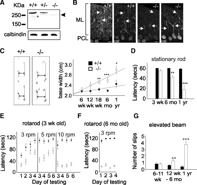
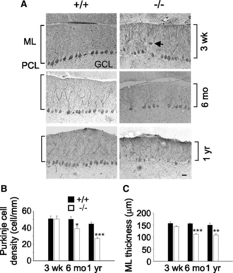
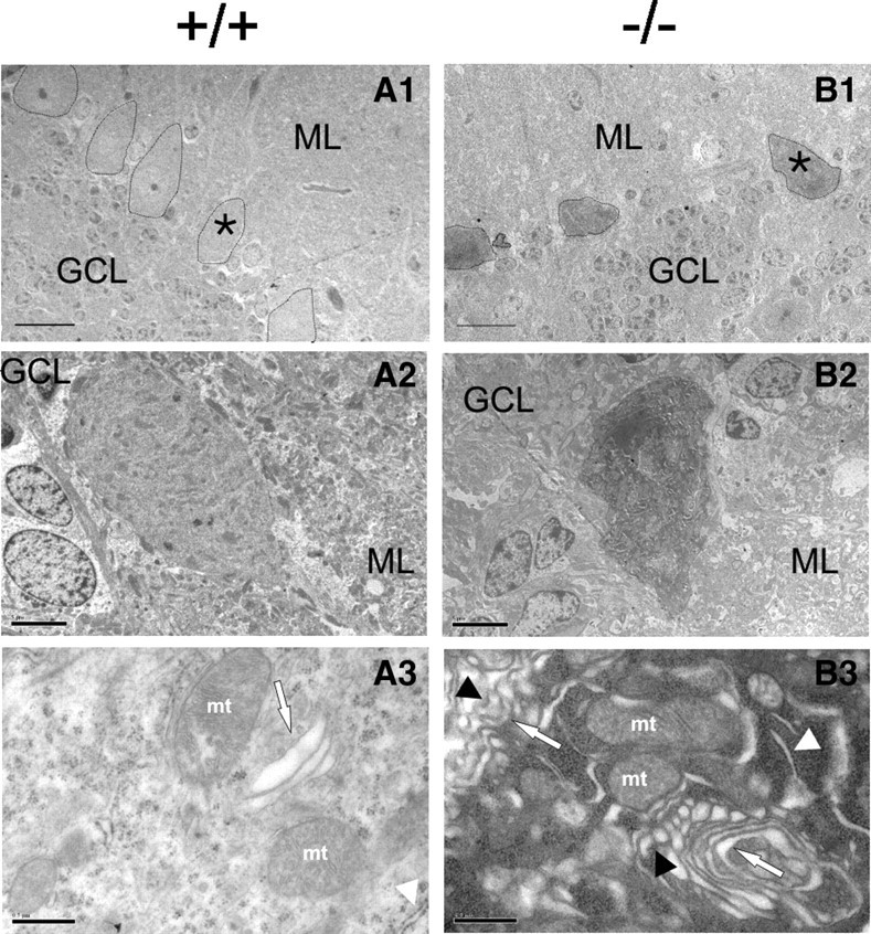
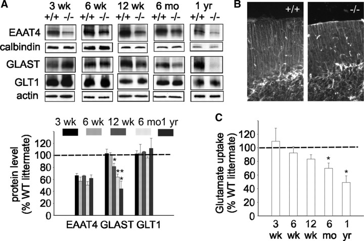
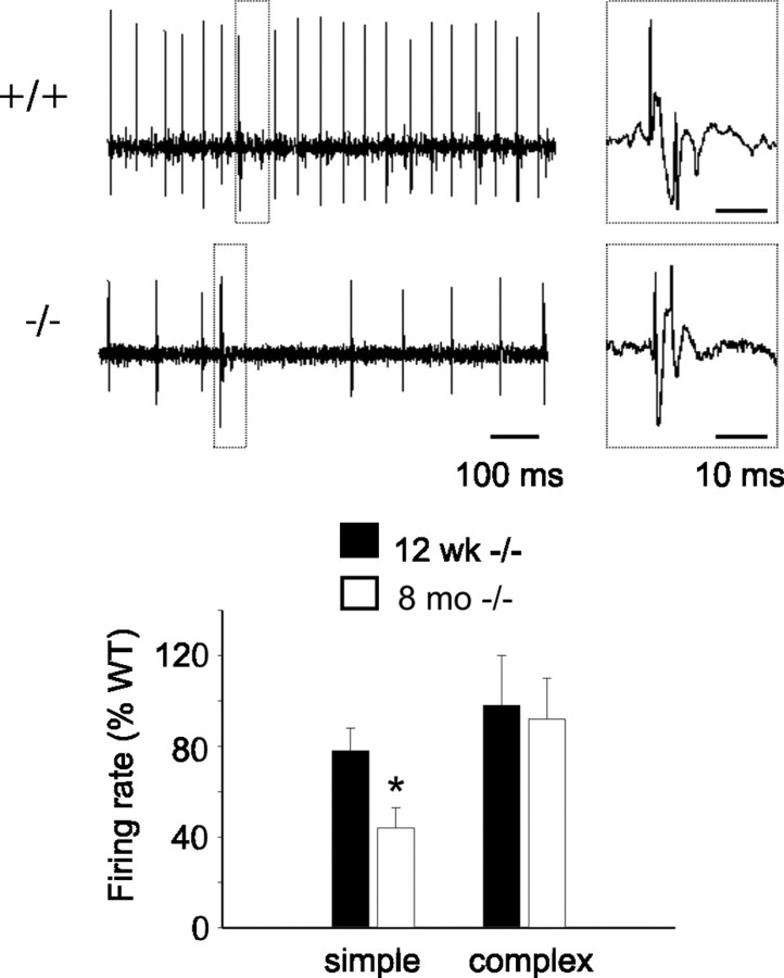
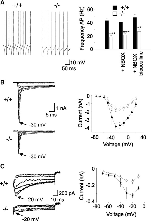
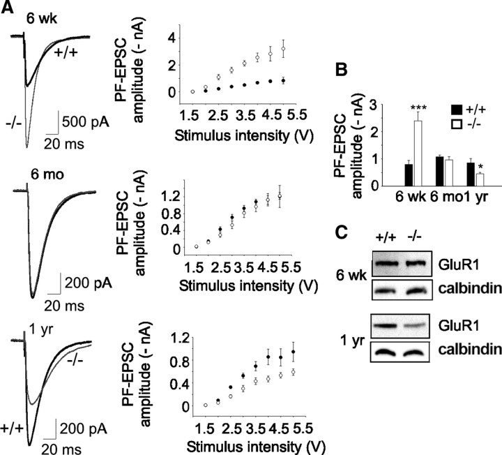
Mutations in SPTBN2, the gene encoding beta-III spectrin, cause spinocerebellar ataxia type 5 in humans (SCA5), a neurodegenerative disorder resulting in loss of motor coordination. How these mutations give rise to progressive ataxia and what the precise role beta-III spectrin plays in normal cerebellar physiology are unknown. We developed a mouse lacking full-length beta-III spectrin and found that homozygous mice reproduced features of SCA5 including gait abnormalities, tremor, deteriorating motor coordination, Purkinje cell loss, and cerebellar atrophy (molecular layer thinning). In vivo analysis reveals an age-related reduction in simple spike firing rate in surviving beta-III(-/-) Purkinje cells, whereas in vitro studies show these neurons to have reduced spontaneous firing, smaller sodium currents, and dysregulation of glutamatergic neurotransmission. Our data suggest an early loss of EAAT4- (protein interactor of beta-III spectrin) and a subsequent loss of GLAST-mediated uptake may play a role in neuronal pathology. These findings implicate a loss of beta-III spectrin function in SCA5 pathogenesis and indicate that there are at least two physiological effects of beta-III spectrin loss that underpin a progressive loss of inhibitory cerebellar output, namely an intrinsic Purkinje cell membrane defect due to reduced sodium currents and alterations in glutamate signaling.


Figures

Figure 1.
Figure 1.
Progressive motor impairment in β-III−/− mice. A, Western blot analysis of whole cerebellar homogenates (10 μg) confirms full-length β-III spectrin (270 kDa) is absent in β-III−/− mice, but a smaller molecular weight protein (∼250 kDa; arrowhead) is expressed at low levels in β-III+/− and β-III−/− animals. Degradation product is identified by an asterisk. Calbindin levels confirm equal protein loading. B, Cerebellar sections, from 3-week-old mice, immunostained with anti-β-III spectrin and anti-calbindin antibody show intense β-III labeling (arrow) of WT (+/+) dendritic tree but faint staining in β-III−/− mice, although still present in proximal and distal dendrites. ML, Molecular layer; PCL, Purkinje cell layer. Scale bar, 50 μm. C, Left, Representative footprints of 18-week-old WT and β-III−/− littermates. Base width shown by double arrow and stride length by solid line. Right, Summary data showing significant increase in β-III−/− base width compared with WT at 18 weeks (p = 0.0078), 6 months (p = 2.82 × 10⁻⁶), and 1 year (p = 0.02) of age. D, Time mice remained on stationary rod before falling reveals 6-month-old and 1-year-old β-III−/− mice are impaired (p = 0.004 and 0.0007). Mice were given four consecutive trials, with maximum time of 60 s. E, Latency of 3-week-old animals to fall from rotarod at 3, 5, and 10 rpm. Mice were given four trials per day and allowed a maximum retention time of 120 s per trial. Both genotypes improved performance during consecutive days at 3 and 5 rpm, but difference in latency to fall remained significant in all comparisons, except on day 3 at 3 rpm (p = 0.218). F, 6-month-old β-III−/− mice failed to stay on rotarod at 3 rpm and showed no improvement. G, Number of hindlimb slips β-III−/− mice made when crossing narrow, elevated beam increased from 12 weeks of age (p = 0.001 and 0.0006). All data are means ± SEM (WT, N = 5–12; β-III−/−, N = 4–15). *p < 0.05; **p < 0.01; ***p < 0.001.
Figure 2.
Figure 2.
Cerebellar degeneration in old β-III−/− spectrin mice. A, Cerebellar sections immunostained with anti-calbindin D antibody reveal some ectopically expressed Purkinje cells (arrow) in 3-week-old β-III−/− mice and shrunken Purkinje cell soma in 6-month-old and 1-year-old β-III−/− mice. ML, Molecular layer; GCL, granule cell layer; PCL, Purkinje cell layer. Scale bar, 50 μm. B, Mean Purkinje cell density measured from cerebellar folia II-IV, VI, and VIII shows Purkinje cell loss in 6-month-old and 1-year-old but not 3-week-old β-III−/− mice. C, Mean molecular layer thickness shows thinning of molecular layer in old β-III−/− mice. All data are means ± SEM. *p < 0.05; **p < 0.01; ***p < 0.001.
Figure 3.
Figure 3.
Signs of glutamate-mediated excitotoxicity in β-III−/− Purkinje cells. A1–B2, Ultrastructural analysis of cerebellar sections from 6-month-old mice shows normal cell bodies in WT (A1, A2) but shrunken and electron dense soma in β-III−/− (B1, B2). A1, B1, 170× magnification; scale bar, 20 μm. A2, B2, 750× magnification; scale bar, 5 μm. A3, B3, Electron micrographs (8400× magnification; scale bars, 0.5 μm) show dilated smooth endoplasmic reticulum (ER) (B3, white arrowhead), and fragmented Golgi cisternae (arrow) with increased number of vesicles, some of which are invaginated (black arrowhead) in β-III−/− Purkinje cell somas compared with abundant rough ER (A3, white arrowhead) and normal Golgi apparatus (arrow) in WT. ML, Molecular layer; GCL, granule cell layer; mt, mitochondria; all Purkinje cell bodies outlined. *Denotes from which cell high-magnification images were obtained. Note different cell orientation in A2 and B2 compared with A1 and B1.
Figure 4.
Figure 4.
Loss of neuronal and astroglial glutamate transporters in β-III−/− mice. A, Top, Representative Western blots illustrating loss of EAAT4 protein in β-III−/− mice from 3 weeks of age. Loss of astroglial glutamate transporter GLAST observed in β-III−/− mice from 12 weeks of age. No loss of GLT1 seen at any age. Bottom, Densitometry data quantifying levels. EAAT4 levels normalized with calbindin, a Purkinje cell-specific marker (p < 0.02 for all ages). GLAST and GLT1 normalized with actin. B, Cerebellar sections immunostained with anti-GFAP antibody show no astrogliosis in 1-year-old β-III−/− mice. C, Glutamate uptake assays on whole cerebellar homogenates show reduced uptake in β-III−/− mice from 12 weeks of age. All data are means ± SEM (N = 3–6). *p < 0.05; **p < 0.01.
Figure 5.
Figure 5.
Reduced in vivo simple spike firing rate in old β-III−/− mice compared with young β-III−/− mice. Top, Representative trace of in vivo Purkinje cell output from 8-month-old WT and β-III−/− animals. Complex spike (dashed box) enlarged on right of trace. Bottom, Firing rates of 12-week-old and 8-month-old β-III−/− mice expressed as percentage of wild-type frequency show reduction in simple but not complex spikes with age. All data are means ± SEM (12 weeks: WT, N = 3, n = 11; β-III−/−, N = 3, n = 12; 8 months: WT, N = 3, n = 8; β-III−/−, N = 3, n = 10; p = 0.02).
Figure 6.
Figure 6.
Reduced spontaneous firing due to smaller sodium currents in Purkinje cells from β-III−/− mice. A, Left, Representative traces of Purkinje cell spontaneous firing measured from 3-week-old WT and β-III−/− littermates. Right, Quantification of mean firing frequency shows a reduction in spike frequency in Purkinje cells from 3-week-old β-III−/− mice compared with WT mice. Presence of NBQX or bicuculline does not abolish genotype difference in firing rate. B, Left, Sodium current traces from representative cells evoked with a series of 50 ms depolarizations from a holding potential of −90 mV to potentials ranging from −80 to + 20 mV in 10 mV increments. Right, Current–voltage relationship shows reduced whole-cell current in β-III−/− mice. C, Left, Representative traces of resurgent sodium currents in WT and β-III−/− mice. Currents evoked using a 20 ms step to + 30 mV, followed by repolarizations from –20 to –70 mV. Right, Mean peak resurgent sodium current versus voltage shows reduced resurgent current in β-III−/− mice. All data are means ± SEM. **p < 0.01; ***p < 0.001.
Figure 7.
Figure 7.
Altered PF-EPSCs in β-III−/− mice. A, Left, Representative EPSC waveforms (4 V stimulus) from 6-week-old, 6-month-old, and 1-year-old WT and β-III−/− littermates. Right, Mean PF-EPSC amplitudes versus stimulus intensity shows consistent differences between WT and β-III−/− cells at different stimuli. B, Mean peak amplitude of EPSCs at 4 V stimulus shows changes in PF-evoked currents with age in β-III−/− cells (6 weeks: N = 3, n = 10, p = 0.001; 6 months: WT, N = 2, n = 10; β-III−/−, N = 2, n = 9, p = 0.44; 1 year: WT, N = 1, n = 6; β-III−/−, N = 2, n = 11, p = 0.05). C, Western blot analysis shows loss of GluR1 in 1-year-old but no change in 6-week-old β-III−/− mice.

Similar articles

Cited by

References

    1. Akemann W, Knöpfel T. Interaction of Kv3 potassium channels and resurgent sodium current influences the rate of spontaneous firing of Purkinje neurons. J Neurosci. 2006;26:4602–4612. - PMC - PubMed
    1. Baines AJ. Evolution of spectrin function in cytoskeletal and membrane networks. Biochem Soc Trans. 2009;37:796–803. - PubMed
    1. Browne DL, Gancher ST, Nutt JG, Brunt ER, Smith EA, Kramer P, Litt M. Episodic ataxia/myokymia syndrome is associated with point mutations in the human potassium channel gene, KCNA1. Nat Genet. 1994;8:136–140. - PubMed
    1. Bürk K, Zühlke C, König IR, Ziegler A, Schwinger E, Globas C, Dichgans J, Hellenbroich Y. Spinocerebellar ataxia type 5: clinical and molecular genetic features of a German kindred. Neurology. 2004;62:327–329. - PubMed
    1. Burright EN, Clark HB, Servadio A, Matilla T, Feddersen RM, Yunis WS, Duvick LA, Zoghbi HY, Orr HT. SCA1 transgenic mice: a model for neurodegeneration caused by an expanded CAG trinucleotide repeat. Cell. 1995;82:937–948. - PubMed

Publication types

MeSH terms

LinkOut - more resources