Comparative Study
. 2010 Apr 7;30(14):4868-76.
doi: 10.1523/JNEUROSCI.5951-09.2010.

In vivo deletion of immunoglobulin domains 5 and 6 in neurofascin (Nfasc) reveals domain-specific requirements in myelinated axons

Affiliations
Comparative Study

In vivo deletion of immunoglobulin domains 5 and 6 in neurofascin (Nfasc) reveals domain-specific requirements in myelinated axons

Courtney Thaxton et al. J Neurosci. .

Abstract

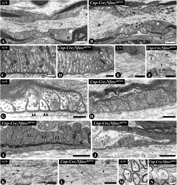
The formation of paranodal axo-glial junctions is critical for the rapid and efficient propagation of nerve impulses. Genetic ablation of genes encoding the critical paranodal proteins Caspr, contactin (Cont), and the myelinating glia-specific isoform of Neurofascin (Nfasc(NF155)) results in the disruption of the paranodal axo-glial junctions, loss of ion channel segregation, and impaired nerve conduction, but the mechanisms regulating their interactions remain elusive. Here, we report that loss of immunoglobulin (Ig) domains 5 and 6 in Nfasc(NF155) in mice phenocopies complete ablation of Nfasc(NF155). The mutant mice lack paranodal septate junctions, resulting in the diffusion of Caspr and Cont from the paranodes, and redistribution of the juxtaparanodal potassium channels toward the nodes. Although critical for Nfasc(NF155) function, we find that Ig5-6 are dispensable for nodal Nfasc(NF186) function. Moreover, in vitro binding assays using Ig5-6 deletion constructs reveal their importance for the association of Nfasc(NF155) with Cont. These findings provide the first molecular evidence demonstrating domain-specific requirements controlling the association of the paranodal tripartite complex in vivo. Our studies further emphasize that in vivo structure/function analysis is necessary to define the unique protein-protein interactions that differentially regulate the functions of Neurofascins during axonal domain organization.


Figures

Figure 1.
Figure 1.
Genetic ablation of Ig5-6 from Nfasc. A, Schematic diagram illustrating Nfasc wild-type and NfascΔIg5-6 isoforms. B, Generation of the conditional NfascIgFlox and NfascΔIg5-6 alleles. C, PCR amplification of genomic DNA for identifying wild-type (+/+), heterozygous (+/NfascIgFlox), and homozygous floxed (NfascIgFlox) Nfasc alleles, and Cre, in Cnp-Cre;NfascIgFlox and β-Act-Cre;NfascIgFlox mutant mice. D, Photographic display of the wild-type (+/+) and NfascNF155ΔIg5-6 (Cnp-Cre; NfascIgFlox) mice. E, Immunoblot analysis of wild-type (+/+) and NfascNF155ΔIg5-6 (Cnp-Cre; NfascIgFlox) spinal cord lysates against the antibodies indicated. F, Immunoblot analysis of wild-type (+/+), heterozygous (+/β-Act-Cre;NfascIgFlox), and NfascΔIg5-6 (β-Act-Cre;NfascIgFlox) spinal cord lysates against the indicated antibodies. The asterisks denote Ig5-6-specific deleted isoforms.
Figure 2.
Figure 2.
Loss of Ig5-6 in NfascNF155 results in axonal domain disorganization. AR, Sciatic nerve fibers from (A–H) P20 NfascNF155ΔIg5-6 (Cnp-Cre;NfascIgFlox) and (I–N) P18 NfascΔIg5-6 (β-Act-Cre;NfascIgFlox) mice with age-matched wild-type (+/+) littermates and cervical spinal cord white matter tracts from (O–R) P20 wild-type (+/+) and NfascNF155ΔIg5-6 (Cnp-Cre;NfascIgFlox) mice were immunostained with a host of antibodies against juxtaparanodal, paranodal, and nodal proteins. All images were collected using identical confocal settings as the control and are representative of the three independent experiments.
Figure 3.
Figure 3.
NfascNF155ΔIg5-6 mutant mice fail to establish paranodal axo-glial junctions. Wild-type (+/+) and NfascNF155ΔIg5-6 (Cnp-Cre; NfascIgFlox) myelinated axons from P20 mice were analyzed by electron microscopy. AL, Longitudinal sections were taken of PNS sciatic nerve fibers (A–H) and spinal cord white matter tracts (I–L). M, N, Cross sections of Purkinje axons within the cerebellar white matter. Arrows in F denote organelle accumulation. Arrowheads in G highlight the electron-dense paranodal septate-like junctions. Arrowheads in K–L mark the arrays of neurofilaments of the axonal cytoskeleton. Asterisks in J indicate the infiltrating astrocytic process, and in M–N they mark axons of interest. EL indicates everted myelin loops. Scale bars represent 0.2 μm (C, D, GJ), 0.5 μm (E, F, K, L), and 1 μm (A, B, M, N).
Figure 4.
Figure 4.
Ig5-6 are essential for NfascNF155 association with Cont. A, B, Immunoblot analysis of transiently transfected HEK293 (A) cells expressing Fc-tagged wild-type NfascNF155 (NF155-Fc), NfascNF155lacking Ig5-6 (NF155ΔIg5/6-Fc), and Glmdn (Glmdn-Fc); and CHO-Lec1 (B) cells transiently expressing HA-tagged wild-type NfascNF186 (NF186-HA), and NfascNF186 deleted of Ig5-6 (NF186ΔIg5/6-HA). C, D, In vitro pull-down assays of CHO-Lec1 cells transiently expressing Cont (C) or rat brain lysates (D) against Fc alone, wild-type NfascNF155(WT-Fc), and Ig5-6 deleted NfascNF155 (ΔIg-Fc). E, F, Immunobinding assays of CHO-Lec1 cells transiently expressing Cont alone, Cont with Caspr (Cont + Caspr), or Glmdn (Glmdn-TM) exposed to NF155-Fc (E), or NF155ΔIg5/6-Fc (F). G, Immunostaining of CHO-Lec1 cells transiently expressing NF186-HA and NF186ΔIg5/6-HA and binding assays Glmdn-Fc. Phase contrast images of the transfected cells (in E, F, G) indicate the health, numbers, and viability of the cells.

Similar articles

Cited by

References

    1. Banerjee S, Bhat MA. Glial ensheathment of peripheral axons in Drosophila. J Neurosci Res. 2008;86:1189–1198. - PMC - PubMed
    1. Bhat MA, Rios JC, Lu Y, Garcia-Fresco GP, Ching W, St Martin M, Li J, Einheber S, Chesler M, Rosenbluth J, Salzer JL, Bellen HJ. Axon-glia interactions and the domain organization of myelinated axons requires neurexin IV/Caspr/Paranodin. Neuron. 2001;30:369–383. - PubMed
    1. Bonnon C, Bel C, Goutebroze L, Maigret B, Girault JA, Faivre-Sarrailh C. PGY repeats and N-glycans govern the trafficking of paranodin and its selective association with contactin and neurofascin-155. Mol Biol Cell. 2007;18:229–241. - PMC - PubMed
    1. Boyle ME, Berglund EO, Murai KK, Weber L, Peles E, Ranscht B. Contactin orchestrates assembly of the septate-like junctions at the paranode in myelinated peripheral nerve. Neuron. 2001;30:385–397. - PubMed
    1. Charles P, Tait S, Faivre-Sarrailh C, Barbin G, Gunn-Moore F, Denisenko-Nehrbass N, Guennoc AM, Girault JA, Brophy PJ, Lubetzki C. Neurofascin is a glial receptor for the paranodin/Caspr-contactin axonal complex at the axoglial junction. Curr Biol. 2002;12:217–220. - PubMed

Publication types

MeSH terms