

Quantification of gammaH2AX foci in response to ionising radiation
- PMID: 20372103
- PMCID: PMC3164074
- DOI: 10.3791/1957
Quantification of gammaH2AX foci in response to ionising radiation
Abstract
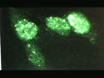
DNA double-strand breaks (DSBs), which are induced by either endogenous metabolic processes or by exogenous sources, are one of the most critical DNA lesions with respect to survival and preservation of genomic integrity. An early response to the induction of DSBs is phosphorylation of the H2A histone variant, H2AX, at the serine-139 residue, in the highly conserved C-terminal SQEY motif, forming gammaH2AX(1). Following induction of DSBs, H2AX is rapidly phosphorylated by the phosphatidyl-inosito 3-kinase (PIKK) family of proteins, ataxia telangiectasia mutated (ATM), DNA-protein kinase catalytic subunit and ATM and RAD3-related (ATR)(2). Typically, only a few base-pairs (bp) are implicated in a DSB, however, there is significant signal amplification, given the importance of chromatin modifications in DNA damage signalling and repair. Phosphorylation of H2AX mediated predominantly by ATM spreads to adjacent areas of chromatin, affecting approximately 0.03% of total cellular H2AX per DSB(2,3). This corresponds to phosphorylation of approximately 2000 H2AX molecules spanning approximately 2 Mbp regions of chromatin surrounding the site of the DSB and results in the formation of discrete gammaH2AX foci which can be easily visualized and quantitated by immunofluorescence microscopy(2). The loss of gammaH2AX at DSB reflects repair, however, there is some controversy as to what defines complete repair of DSBs; it has been proposed that rejoining of both strands of DNA is adequate however, it has also been suggested that re-instatement of the original chromatin state of compaction is necessary(4-8). The disappearence of gammaH2AX involves at least in part, dephosphorylation by phosphatases, phosphatase 2A and phosphatase 4C(5,6). Further, removal of gammaH2AX by redistribution involving histone exchange with H2A.Z has been implicated(7,8). Importantly, the quantitative analysis of gammaH2AX foci has led to a wide range of applications in medical and nuclear research. Here, we demonstrate the most commonly used immunofluorescence method for evaluation of initial DNA damage by detection and quantitation of gammaH2AX foci in gamma-irradiated adherent human keratinocytes(9).
Similar articles
-
Evaluation of the spatial distribution of gammaH2AX following ionizing radiation.J Vis Exp. 2010 Aug 7;(42):2203. doi: 10.3791/2203. J Vis Exp. 2010. PMID: 20736911 Free PMC article.
-
Quantitation of gammaH2AX foci in tissue samples.J Vis Exp. 2010 Jun 28;(40):2063. doi: 10.3791/2063. J Vis Exp. 2010. PMID: 20613712 Free PMC article.
-
gammaH2AX foci form preferentially in euchromatin after ionising-radiation.PLoS One. 2007 Oct 24;2(10):e1057. doi: 10.1371/journal.pone.0001057. PLoS One. 2007. PMID: 17957241 Free PMC article.
-
Mechanism of elimination of phosphorylated histone H2AX from chromatin after repair of DNA double-strand breaks.Mutat Res. 2010 Mar 1;685(1-2):54-60. doi: 10.1016/j.mrfmmm.2009.08.001. Epub 2009 Aug 12. Mutat Res. 2010. PMID: 19682466 Review.
-
Gamma-H2AX - a novel biomarker for DNA double-strand breaks.In Vivo. 2008 May-Jun;22(3):305-9. In Vivo. 2008. PMID: 18610740 Review.
Cited by
-
Sperm DNA fragmentation testing in clinical management of reproductive medicine.Reprod Med Biol. 2023 Oct 31;22(1):e12547. doi: 10.1002/rmb2.12547. eCollection 2023 Jan-Dec. Reprod Med Biol. 2023. PMID: 37915974 Free PMC article. Review.
-
Non-linear dose response of DNA double strand breaks in response to chronic low dose radiation in individuals from high level natural radiation areas of Kerala coast.Genes Environ. 2023 May 1;45(1):16. doi: 10.1186/s41021-023-00273-6. Genes Environ. 2023. PMID: 37127760 Free PMC article.
-
Trichostatin A accentuates doxorubicin-induced hypertrophy in cardiac myocytes.Aging (Albany NY). 2010 Oct;2(10):659-68. doi: 10.18632/aging.100203. Aging (Albany NY). 2010. PMID: 20930262 Free PMC article.
-
Functional interrogation of adult hypothalamic neurogenesis with focal radiological inhibition.J Vis Exp. 2013 Nov 14;(81):e50716. doi: 10.3791/50716. J Vis Exp. 2013. PMID: 24300415 Free PMC article.
-
Robo1 and vimentin regulate radiation-induced motility of human glioblastoma cells.PLoS One. 2018 Jun 4;13(6):e0198508. doi: 10.1371/journal.pone.0198508. eCollection 2018. PLoS One. 2018. PMID: 29864155 Free PMC article.
References
Publication types
MeSH terms
Substances
LinkOut - more resources
Full Text Sources
Research Materials
Miscellaneous
