. 2010 May;36(5):1299-307.
doi: 10.3892/ijo_00000615.

Inhibition of NF-kappaB and Akt pathways by an antibody-avidin fusion protein sensitizes malignant B-cells to cisplatin-induced apoptosis

Affiliations

Inhibition of NF-kappaB and Akt pathways by an antibody-avidin fusion protein sensitizes malignant B-cells to cisplatin-induced apoptosis

Eriko Suzuki et al. Int J Oncol. 2010 May.

Abstract

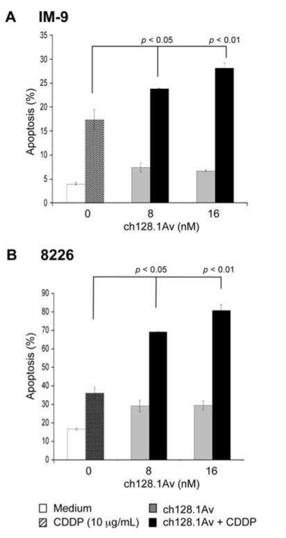
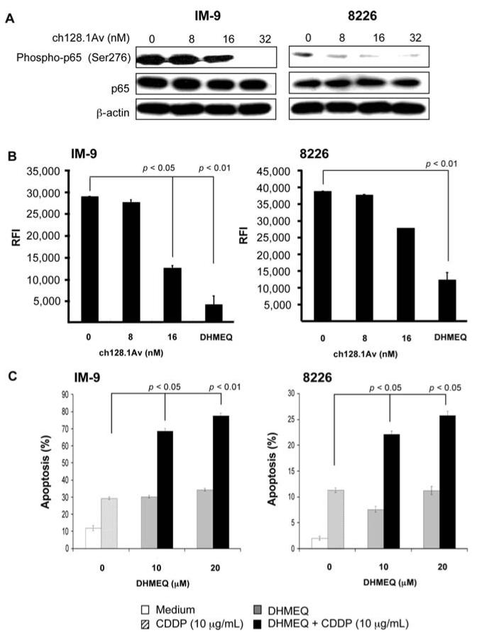
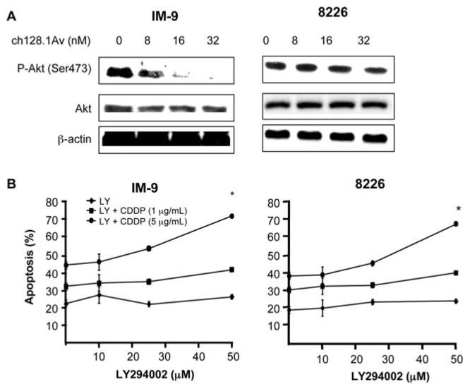
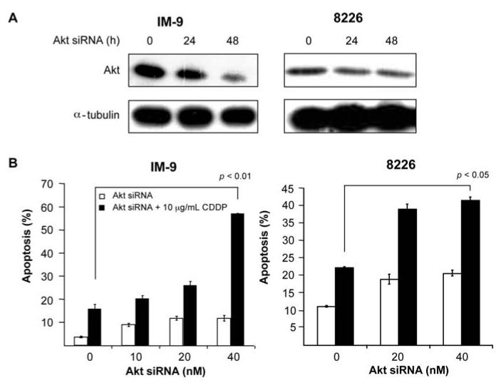
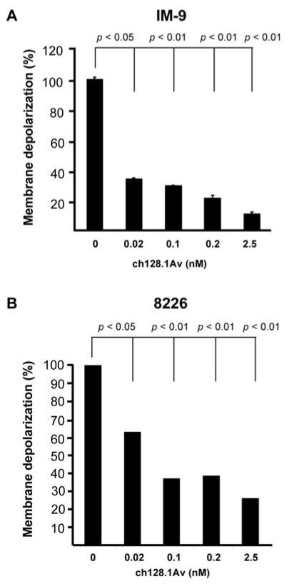
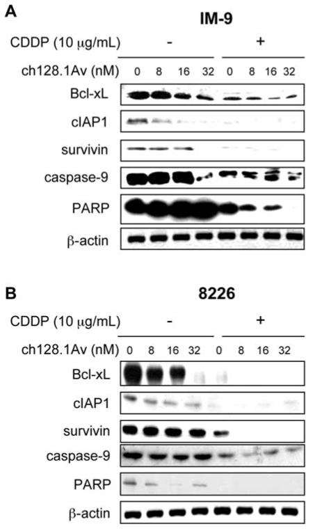
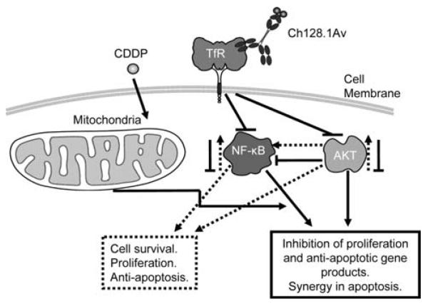
Multiple myeloma (MM) is an incurable disease of malignant plasma cells. Recent therapeutic advancements have resulted in improved response rates, however, there is no improvement in overall survival, therefore, new therapeutics are needed. Since the transferrin receptor is upregulated on the surface of MM cells, we previously developed an antibody fusion protein consisting of an IgG3 specific for the human transferrin receptor 1 (TfR1, CD71) genetically fused to avidin at its carboxy-terminus (ch128.1Av). We have previously shown that ch128.1Av exhibits intrinsic cytotoxicity against certain malignant B-cells by disrupting the cycling of the TfR and decreasing TfR cell surface expression resulting in lethal iron starvation. In addition, ch128.1Av can sensitize malignant cells to apoptosis induced by gambogic acid, a herbal drug used in Chinese medicine. In this study, we hypothesized that ch128.1Av may also sensitize drug-resistant malignant B-cells to chemotherapeutic agents by inhibiting key survival pathways. In this study we show that ch128.1Av sensitizes malignant B-cells to apoptosis induced by cisplatin (CDDP). The sensitization by ch128.1Av resulted in the inhibition of the constitutively activated Akt and NF-kappaB survival/antiapoptotic pathways and downstream decreased expression of antiapoptotic gene products such as BclxL and survivin. The direct role of the inhibition of the Akt and NF-kappaB pathways by ch128.1Av in CDDP-mediated cytotoxicity was demonstrated by the use of specific chemical inhibitors and siRNA which mimicked the effects of ch128.1Av. Overall, this study provides evidence of the therapeutic potential of ch128.1Av as a chemo-sensitizing agent in drug-resistant tumor cells.


Figures

Figure 1
Figure 1
ch128.1Av sensitizes tumor cells to CDDP-induced apotosis. IM-9 (A) and 8226 cells (B) were pre-treated with various concentrations of ch128.1Av for 24 h followed by treatment with CDDP (10 μg/ml) for an additional 24 h. Apoptosis was determined using the PI/Annexin-V as described in methods. The data represent the mean values ±SD from 3 independent experiments. p-value: cells treated with a single agent (ch128.1Av or CDDP) vs. combined treatment with CDDP (Student's t-test).
Figure 2
Figure 2
Inhibition of NF-κB by ch128.1Av and the role of NF-κB in tumor cell sensitization to CDDP-induced apoptosis. (A) ch128.1Av inhibits NF-κB activation as evidenced by the decreased level of phospho-p65 in IM-9 (left) and 8226 (right) cells. Cells were treated with 0-32 nM of ch128.1Av for 24 h and cell lysates were prepared and examined by Western blot analysis for phospho-p65, p65 and β-actin expression. Blots are representative of 3 independent but reproducible experiments. (B) NF-κB DNA-binding activity in IM-9 (left) and 8226 cells (right). Cells were treated with 0-32 nM of ch128.1Av for 24 h and cell lysates were prepared and examined for DNA-binding analysis using Trans AM DNA-binding analysis kit as described in Materials and methods. The NF-κB inhibitor, DHMEQ, was used as an internal positive control. p-value: medium control compared to treatment with 16 nM of ch128.1Av (Student's t-test). (C) IM-9 (left) and 8226 cells (right) were treated with various concentrations of (-)-DHMEQ (0-20 μg/ml) for 2 h and treated with CDDP (10 μg/ml) for an additional 24 h. Apoptosis was determined using the PI/Annexin-V method. p-value: single (-)-DHMEQ or CDDP treatment vs. combined treatment (Student's t-test).
Figure 3
Figure 3
Inhibition of Akt activity by ch128.1Av and the direct role of Akt inhibition via LY294002 in the sensitization to CDDP-induced apoptosis. (A) The ch128.1Av inhibits phospho-Akt in IM-9 cells (left) and 8226 cells (right). Cells were treated with 0-32 nM of ch128.1Av for 24 h and cell lysates were prepared and examined by Western blot analysis for phospho-Akt, Akt and β-actin. Blots are representative of 3 independent but reproducible experiments. (B) Treatment of IM-9 cells (left) and 8226 cells (right) with a specific Akt inhibitor LY294002 results in sensitization to CDDP-induced apoptosis. IM-9 and 8226 cells were treated with various concentrations of CDDP (1 or 5 μg/ml) for 24 h in the presence or absence of LY294002 (0-50 μM) and apoptosis was determined using the PI/Annexin-V method. *p-value: single cell treatment with LY294002 or CDDP vs. combinational treatment. (Student's t-test).
Figure 4
Figure 4
Direct inhibition of Akt by siRNA in cells results in sensitization to CDDP-induced apoptosis. (A) IM-9 (left) or 8226 (right) cells were treated with 8 nM of either Akt siRNA or control scramble siRNA for 0-48 h. Cell lysates were examined for total Akt levels by Western blot analysis. α-tubulin served as an internal control for loading. (B) Cells were treated with increasing concentrations of Akt siRNA for 48 h and CDDP (10 μg/ml) for an additional 24 h, and apoptosis was determined using the PI/Annexin-V method. *p-value: single Akt siRNA or CDDP treatment vs. combined treatment (Student's t-test).
Figure 5
Figure 5
ch128.1Av-induces mitochondorial membrane potential depolarization in malignant B-cells. Mitochondorial membrane depolarization was assessed in IM-9 cells (A) and 8226 cells (B) using the DiOC6 dye by flow cytometry as described in Materials and methods. The cells were treated with various concentrations of ch128.1Av (0.02-2.5 nM) for 24 h and then examined for membrane potential. Values are expressed as Mean Fluorescence Intensity (MFI) of DiOC6 incorporation. Untreated cells are represented as 100 and treated cells are calculated as percentage of untreated cells. The data represent the mean ± SD of 3 independent experiments. p-value: medium control vs. treatment with 16 nM of ch128.1Av (Student's t-test).
Figure 6
Figure 6
ch128.1Av sensitizes malignant B-cells to apoptosis by CDDP via activation of the intrinsic apoptotic pathway. Total cell lysates derived from IM-9 cells (A) and 8226 cells (B) treated with various concentrations of ch128.1Av (0-32 nM) and CDDP (10 mg/ml) for 24 h were examined for levels of expression for BclxL, cIAP-1, survivin, caspase-9 and PARP by Western analysis. β-actin served as an internal control for loading. The blots are representative of three independent experiments.
Figure 7
Figure 7
Schematic diagram of the mechanism by which ch128.1Av sensitizes malignant B-cells to apoptosis by chemotherapeutic drugs. Malignant B-cells constitutively express NF-κB and Akt activating pathways. These result downstream in the transcription and expression of various gene products that regulate cell survival, proliferation and resistance to apoptosis. The tumor cells overexpress the transferrin receptor. Upon binding of ch128.1Av to its corresponding transferrin receptor, the complex is internalized and several modifications take place in the cancer cells. Both the NF-κB and Akt activating pathways are inhibited and consequently, inhibition of cell survival, proliferation and anti-apoptotic gene products are observed. In addition, ch128.1Av also affects the mitochondria and depolarizes the membrane potential. Likewise, treatment with CDDP also affects the mitochondria, however, the combination treatment results in potentiation of apoptosis in the resistant malignant B-cells.

Similar articles

Cited by

References

    1. Sloan B, Scheinfeld NS. Pazopanib, a VEGF receptor tyrosine kinase inhibitor for cancer therapy. Curr Opin Investig Drugs. 2008;9:1324–1335. - PubMed
    1. Law CL, Gordon KA, Collier J, et al. Preclinical antilymphoma activity of a humanized anti-CD40 monoclonal antibody, SGN-40. Cancer Res. 2005;65:8331–8338. - PubMed
    1. Lee JH, Koo TH, Yoon H, et al. Inhibition of NF-kappa B activation through targeting I kappa B kinase by celastrol, a quinone methide triterpenoid. Biochem Pharmacol. 2006;72:1311–1321. - PubMed
    1. Watanabe M, Dewan MZ, Okamura T, et al. A novel NF-kappaB inhibitor DHMEQ selectively targets constitutive NF-kappaB activity and induces apoptosis of multiple myeloma cells in vitro and in vivo. Int J Cancer. 2005;114:32–38. - PubMed
    1. Tatetsu H, Okuno Y, Nakamura M, et al. Dehydroxymethylepoxyquinomicin, a novel nuclear factor-kappaB inhibitor, induces apoptosis in multiple myeloma cells in an IkappaBalpha-independent manner. Mol Cancer Ther. 2005;4:1114–1120. - PubMed

Publication types

MeSH terms