

Immunohistochemical identification of notochordal markers in cells in the aging human lumbar intervertebral disc
- PMID: 20372940
- PMCID: PMC2989227
- DOI: 10.1007/s00586-010-1392-z
Immunohistochemical identification of notochordal markers in cells in the aging human lumbar intervertebral disc
Abstract
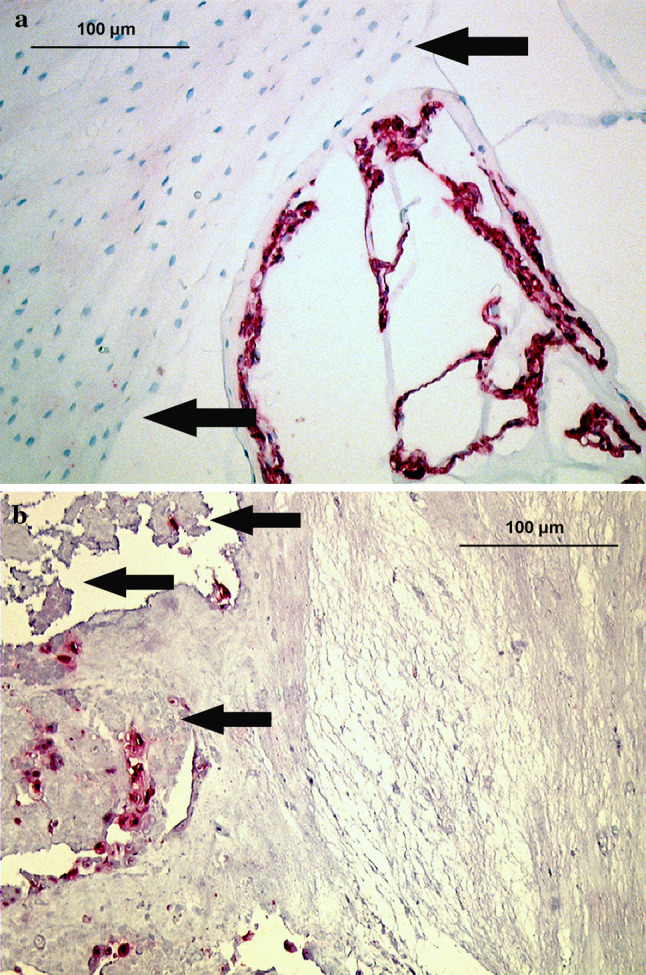
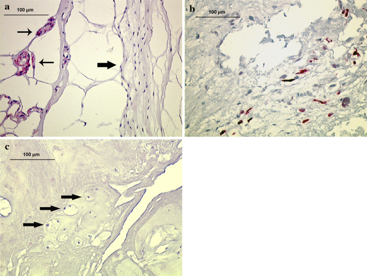
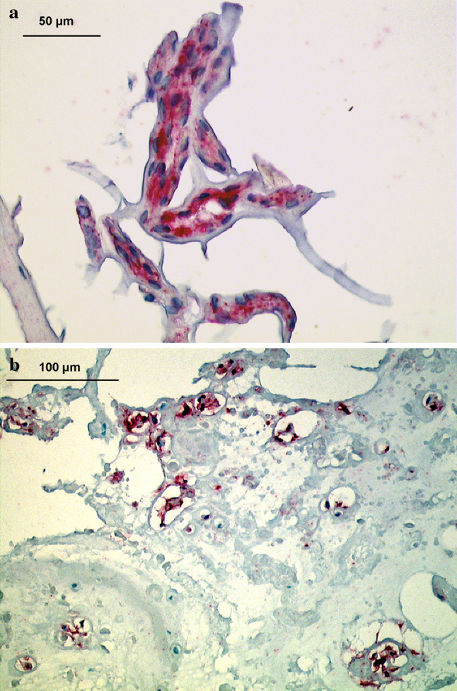
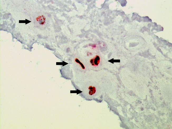
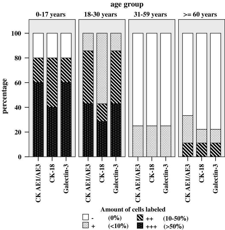
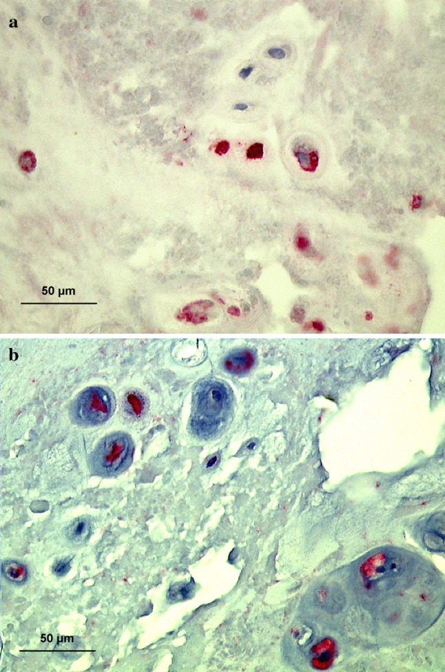
The fate of notochord cells during disc development and aging is still a subject of debate. Cells with the typical notochordal morphology disappear from the disc within the first decade of life. However, the pure morphologic differentiation of notochordal from non-notochordal disc cells can be difficult, prompting the use of cellular markers. Previous reports on these notochordal cell markers only explored the occurrence in young age groups without considering changes during disc degeneration. The aim of this study, therefore, was to investigate presence, localization, and abundance of cells expressing notochordal cell markers in human lumbar discs during disc development and degeneration. Based on pilot studies, cytokeratins CK-8, -18 and -19 as well as Galectin-3 were chosen from a broad panel of potential notochordal cell markers and used for immunohistochemical staining of 30 human lumbar autopsy samples (0-86 years) and 38 human surgical disc samples (26-69 years). In the autopsy group, 80% of fetal to adolescent discs (0-17 years) and 100% of young adult discs (18-30 years) contained many cells with positive labeling. These cells were strongly clustered and nearly exclusively located in areas with granular changes (or other matrix defects), showing predominantly a chondrocytic morphology as well as (in a much lesser extent) a fibrocytic phenotype. In mature discs (31-60 years) and elderly discs (≥ 60 years) only 25 and 22-33%, respectively, contained few stained nuclear cells, mostly associated with matrix defects. In the surgical group, only 16% of samples from young adults (≤ 47 years) exhibited positively labeled cells whereas mature to old surgical discs (>47 years) contained no labeled cells. This is the first study describing the presence and temporo-spatial localization of cells expressing notochordal cell markers in human lumbar intervertebral discs of all ages and variable degree of disc degeneration. Our findings indicate that cells with a (immunohistochemically) notochord-like phenotype are present in a considerable fraction of adult lumbar intervertebral discs. The presence of these cells is associated with distinct features of (early) age-related disc degeneration, particularly with granular matrix changes.
Figures

Similar articles
-
1997 Volvo Award winner in basic science studies. Immunohistologic markers for age-related changes of human lumbar intervertebral discs.Spine (Phila Pa 1976). 1997 Dec 15;22(24):2781-95. doi: 10.1097/00007632-199712150-00001. Spine (Phila Pa 1976). 1997. PMID: 9431614
-
Expression and distribution of tumor necrosis factor alpha in human lumbar intervertebral discs: a study in surgical specimen and autopsy controls.Spine (Phila Pa 1976). 2005 Jan 1;30(1):44-53; discussion 54. doi: 10.1097/01.brs.0000149186.63457.20. Spine (Phila Pa 1976). 2005. PMID: 15626980
-
Tracing notochord-derived cells using a Noto-cre mouse: implications for intervertebral disc development.Dis Model Mech. 2012 Jan;5(1):73-82. doi: 10.1242/dmm.008128. Epub 2011 Oct 25. Dis Model Mech. 2012. PMID: 22028328 Free PMC article.
-
The role of disc cell heterogeneity in determining disc biochemistry: a speculation.Biochem Soc Trans. 2002 Nov;30(Pt 6):839-44. doi: 10.1042/bst0300839. Biochem Soc Trans. 2002. PMID: 12440929 Review.
-
Notochordal cells in the adult intervertebral disc: new perspective on an old question.Crit Rev Eukaryot Gene Expr. 2011;21(1):29-41. doi: 10.1615/critreveukargeneexpr.v21.i1.30. Crit Rev Eukaryot Gene Expr. 2011. PMID: 21967331 Free PMC article. Review.
Cited by
-
Effects of Caffeine on Intervertebral Disc Cell Viability in a Whole Organ Culture Model.Global Spine J. 2022 Jan;12(1):61-69. doi: 10.1177/2192568220948031. Epub 2020 Sep 16. Global Spine J. 2022. PMID: 32935580 Free PMC article.
-
Update on the pathophysiology of degenerative disc disease and new developments in treatment strategies.Open Access J Sports Med. 2010 Oct 14;1:191-9. doi: 10.2147/OAJSM.S9057. Open Access J Sports Med. 2010. PMID: 24198557 Free PMC article. Review.
-
Differential Response of Bovine Mature Nucleus Pulposus and Notochordal Cells to Hydrostatic Pressure and Glucose Restriction.Cartilage. 2020 Apr;11(2):221-233. doi: 10.1177/1947603518775795. Epub 2018 May 29. Cartilage. 2020. PMID: 29808709 Free PMC article.
-
Validation of reference genes in human chordoma.Surg Neurol Int. 2017 Jun 5;8:100. doi: 10.4103/sni.sni_399_16. eCollection 2017. Surg Neurol Int. 2017. PMID: 28695047 Free PMC article.
-
Evoked thalamic neuronal activity following DRG application of two nucleus pulposus derived cell populations: an experimental study in rats.Eur Spine J. 2013 May;22(5):1113-8. doi: 10.1007/s00586-013-2669-9. Epub 2013 Jan 24. Eur Spine J. 2013. PMID: 23341046 Free PMC article.
References
-
- Boos N, Nerlich AG, Wiest I, von der MK, Aebi M (1997) Immunolocalization of type X collagen in human lumbar intervertebral discs during ageing and degeneration. Histochem Cell Biol 108:471–480 - PubMed
Publication types
MeSH terms
LinkOut - more resources
Full Text Sources
Research Materials
