. 2010 Mar;67(3):325-37.
doi: 10.1002/ana.21897.

Triptan-induced latent sensitization: a possible basis for medication overuse headache

Affiliations

Triptan-induced latent sensitization: a possible basis for medication overuse headache

Milena De Felice et al. Ann Neurol. 2010 Mar.

Abstract

Objective: Identification of the neural mechanisms underlying medication overuse headache resulting from triptans.

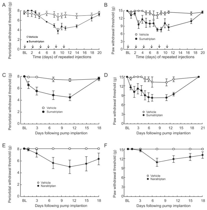
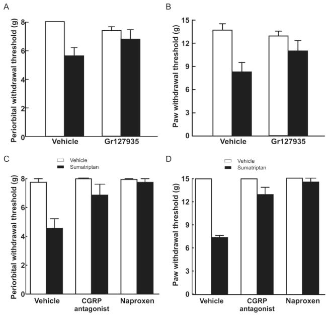
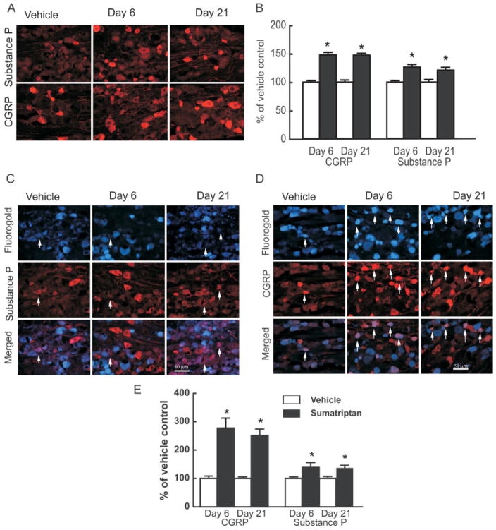
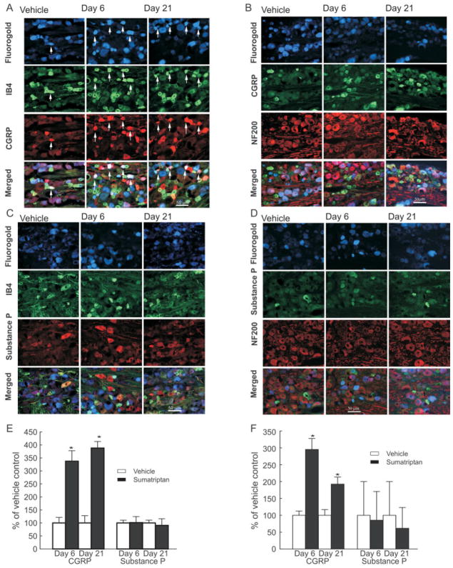
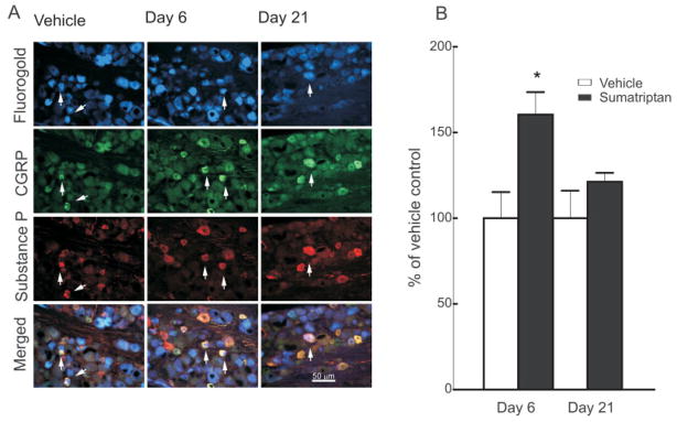
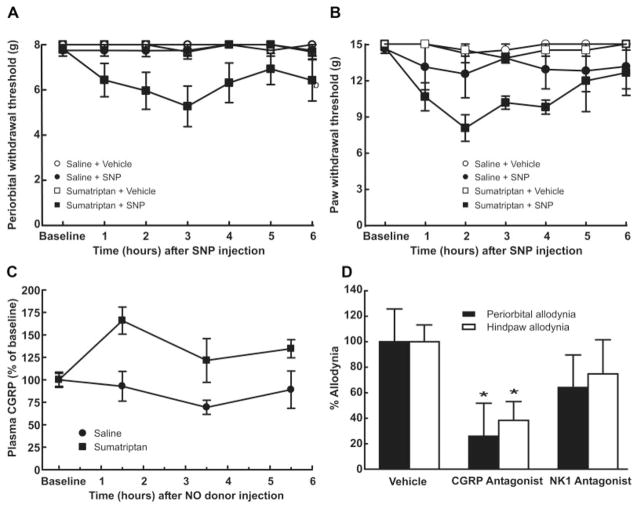
Methods: Triptans were administered systemically to rats by repeated intermittent injections or by continuous infusion over 6 days. Periorbital and hind paw sensory thresholds were measured to detect cutaneous allodynia. Immunofluorescent histochemistry was employed to detect changes in peptidic neurotransmitter expression in identified dural afferents. Enzyme-linked immunoabsorbent assay was used to measure calcitonin gene-related peptide (CGRP) levels in blood.

Results: Sustained or repeated administration of triptans to rats elicited time-dependent and reversible cutaneous tactile allodynia that was maintained throughout and transiently after drug delivery. Triptan administration increased labeling for CGRP in identified trigeminal dural afferents that persisted long after discontinuation of triptan exposure. Two weeks after triptan exposure, when sensory thresholds returned to baseline levels, rats showed enhanced cutaneous allodynia and increased CGRP in the blood following challenge with a nitric oxide donor. Triptan treatment thus induces a state of latent sensitization characterized by persistent pronociceptive neural adaptations in dural afferents and enhanced responses to an established trigger of migraine headache in humans.

Interpretation: Triptans represent the treatment of choice for moderate and severe migraine headaches. However, triptan overuse can lead to an increased frequency of migraine headache. Overuse of these medications could induce neural adaptations that result in a state of latent sensitization, which might increase sensitivity to migraine triggers. The latent sensitization could provide a mechanistic basis for the transformation of migraine to medication overuse headache.


Figures

FIGURE 1
FIGURE 1
Sustained exposure to triptans reduces sensory thresholds to light tactile stimuli applied to the periorbital region and the hind paws of rats. Repeated injections of sumatriptan (0.6mg/kg subcutaneously [sc]) decrease the withdrawal thresholds to light tactile stimuli applied to (A) the periorbital region or (B) the hind paws of rats. Sumatriptan or saline was injected 6 times, at 48-hour intervals, and sensory thresholds were evaluated every 24 hours. Withdrawal responses to von Frey filaments are significantly (p < 0.05) reduced in a time-dependent manner. Continuous infusion of sumatriptan (0.6mg/kg/day sc) decreased withdrawal thresholds to light tactile stimuli applied to (C) the periorbital region or (D) the hind paws of rats. Sumatriptan or saline was continuously administered through an osmotic minipump for 6 days, after which the minipumps were removed. Withdrawal responses to von Frey filaments were significantly (p < 0.05) reduced in a time-dependent manner. Continuous infusion of naratriptan (0.6mg/kg/day sc) decreased withdrawal thresholds to light tactile stimuli applied to (E) the periorbital region or (F) the hind paws of rats. Naratriptan or saline was continuously administered through an osmotic minipump for 6 days, after which the minipumps were removed. Withdrawal responses to von Frey filaments were significantly (p < 0.05) reduced in a time-dependent manner. BL = baseline.
FIGURE 2
FIGURE 2
Effects of sumatriptan exposure are mediated by serotoninergic receptors. Coinfusion of sumatriptan (0.6mg/kg/day) with a nonselective antagonist at 5-hydroxytryptamine1B/1D receptors, GR127935 (1 mg/kg/day), significantly (p < 0.05) prevented the development of both (A) periorbital and (B) hind paw allodynia. There was no significant difference (p > 0.05) between the responses of animals receiving GR127935 with saline and vehicle/saline. Rats received vehicle or sumatriptan through an osmotic minipump for 6 days. On day 6 after evaluation of the cutaneous allodynia in sumatriptan-exposed animals, rats received a single injection of the calcitonin gene-related peptide (CGRP) antagonist α-CGRP(8–37) (0.45mg/kg intravenously), and (C) periorbital and (D) hind paw sensory thresholds were evaluated. The response thresholds of rats receiving α-CGRP(8–37) were significantly (p < 0.05) greater than those of sumatriptan-treated rats receiving vehicle infusion 30 minutes after the injection. Naproxen sodium (100mg/kg subcutaneously) produced a similar significant (p < 0.05) blockade of sumatriptan-induced (C) periorbital and (D) hind paw allodynia within 1 hour of the injection. Neither α-CGRP(8–37) nor naproxen sodium altered the sensory thresholds of saline-treated animals.
FIGURE 3
FIGURE 3
Sustained infusion of sumatriptan (0.6mg/kg/day) for 6 days promoted increased and persistent expression of the neuropeptides calcitonin gene-related peptide (CGRP) and substance P in the trigeminal ganglia of rats. (A) Immunofluorescence labeling for CGRP or substance P in the trigeminal ganglion is shown for sections obtained 6 and 21 days after implantation of a minipump delivering vehicle (saline) or sumatriptan. (B) Sumatriptan exposure resulted in a significant (p < 0.05) increase in CGRP- and substance P-labeled profiles in the trigeminal ganglia, relative to vehicle-infused animals, at both days 6 and 21. Additionally, dural afferents were identified by application of Fluoro-Gold to the dura, and profiles expressing labeling for (C) substance P and (D) CGRP were evaluated in the trigeminal ganglia 6 and 21 days after sumatriptan exposure. (E) The relative proportion of profiles obtained from sumatriptan-treated animals and expressing both the retrograde label and label for CGRP or substance P in the trigeminal ganglia showed a significant and persistent (day 6 and day 21) increase in CGRP and substance P labeling relative to vehicle-infused animals. Notably, the increases in numbers of neuronal profiles labeled for CGRP were significantly (p < 0.05) greater than those expressing substance P. These observations suggest that sumatriptan exposure results in a substantial increase in dural afferents labeled for CGRP, with only a small increase in substance P expression in these cells.
FIGURE 4
FIGURE 4
Sustained infusion of sumatriptan leads to an increased expression of calcitonin gene-related peptide (CGRP) in normally peptide-poor unmyelinated fibers and in myelinated fibers. Dural afferents were identified by administration of Fluoro-Gold to the dura, 4 days before collecting trigeminal tissue for immunofluorescent imaging. Trigeminal ganglion sections were obtained from rats 6 and 21 days after initiation of sumatriptan infusion (0.6mg/kg/day) and labeled for (A, B) CGRP or (C, D) substance P. The sections were also labeled for reactivity to (A, C) IB4 or (B, D) NF200. (E, F) The proportion of retrogradely labeled profiles also showing label for CGRP and for either IB4 or NF200 or for substance P and for either IB4 or NF200 were determined relative to that shown by sections obtained from saline-treated rats. Sumatriptan infusion resulted in a significant (*p < 0.05) increase in retrogradely labeled trigeminal profiles labeled for CGRP and either (E) IB4 or (F) NF200 6 and 21 days after initiation of infusion. In contrast, levels of substance P were not significantly elevated (p > 0.05) in any of the sections studies. Notably, substance P expression in profiles labeled for NF200 was very low, representing <1% of all profiles counted. (F) These low counts resulted in the large standard errors shown. These results indicate that sumatriptan exposure produces a preferential increase in CGRP expression in dural afferents that do not normally express this peptide, or express it at a low level, or in myelinated dural afferents.
FIGURE 5
FIGURE 5
Sumatriptan treatment increased the coexpression of substance P with calcitonin gene-related peptide (CGRP) in dural afferents. (A) After retrolabeling the dural afferents of the trigeminal ganglia, sections were prepared for fluorescent staining to visualize CGRP and substance P at day 6 and 21 after sumatriptan exposure. (B) The numbers of profiles expressing substance P and CGRP were counted. Sumatriptan exposure induced a small but significant (*p < 0.05) increase in coexpression of CGRP and substance P, relative to vehicle-infused animals, observed at day 6 but not at day 21 after sumatriptan pump implantation.
FIGURE 6
FIGURE 6
Sumatriptan-induced latent sensitization. On day 20 after pump implantation, an acute injection of the nitric oxide (NO) donor sodium nitroprusside (SNP) (3 mg/kg intraperitoneally [ip]) significantly (p < 0.05) reduced both the (A) periorbital and (B) hind paw withdrawal thresholds to probing with von Fey filaments. SNP injection in saline-exposed rats did not change the withdrawal thresholds. (C) Rats were pre-exposed to sumatriptan infusion (0.6mg/kg/day for 6 days) and challenged with SNP (3mg/kg ip) after 20 days. Blood samples (1ml) were withdrawn 1.5, 3.5, and 5.5 hours after the injection. Sumatriptan exposure resulted in a significant (p < 0.05) and time-dependent increase in calcitonin gene-related peptide (CGRP) levels. No changes in CGRP blood levels were observed in saline-exposed rats. (D) Twenty days after implantation of minipumps, rats received SNP (3mg/kg ip) followed after 30 minutes by α-CGRP(8–37) (0.45mg/kg intravenously). Systemic administration of α-CGRP(8–37) significantly (*p < 0.05) reduced behavioral signs of periorbital and hind paw tactile allodynia within 1 hour of the injection. In a second set of studies, the NK-1 antagonist L-732,138 (10mg/kg subcutaneously) was administered 30 minutes after SNP. Treatment with L-732,138 failed to abolish the development of tactile allodynia of the face or hind paws. Behavioral responses were not altered by vehicle, α-CGRP(8–37), or L-732,138 in saline exposed rats (data not shown).

Similar articles

Cited by

References

    1. Society HCCotIH. The International Classification of Headache Disorders (second edition) Cephalalgia. 2004;24:1–160. - PubMed
    1. Launer LJ, Terwindt GM, Ferrari MD. The prevalence and characteristics of migraine in a population-based cohort: the GEM study. Neurology. 1999;53:537–542. - PubMed
    1. Bolay H, Moskowitz MA. The neurobiology of migraine and transformation of headache therapy. In: Waxman S, editor. From neuroscience to neurology: neuroscience, molecular medicine, and the therapeutic transformation of neurology. San Diego, CA: Elsevier; 2004. pp. 107–123.
    1. Bolay H, Moskowitz MA. The emerging importance of cortical spreading depression in migraine headache. Rev Neurol (Paris) 2005;161:655–657. - PubMed
    1. Buzzi MG, Bonamini M, Moskowitz MA. Neurogenic model of migraine. Cephalalgia. 1995;15:277–280. - PubMed

MeSH terms