. 2010 Apr;31(4):987-96.
doi: 10.1002/jmri.22112.

Accelerated slice encoding for metal artifact correction

Affiliations

Accelerated slice encoding for metal artifact correction

Brian A Hargreaves et al. J Magn Reson Imaging. 2010 Apr.

Abstract

Purpose: To demonstrate accelerated imaging with both artifact reduction and different contrast mechanisms near metallic implants.

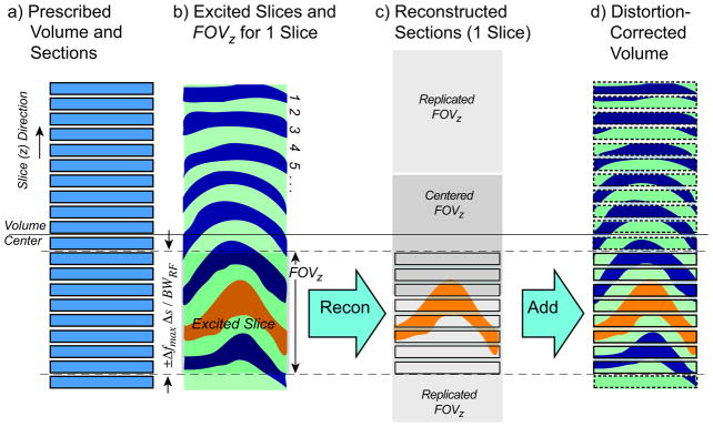
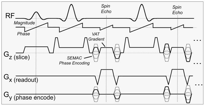
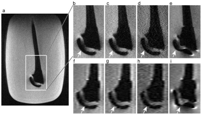
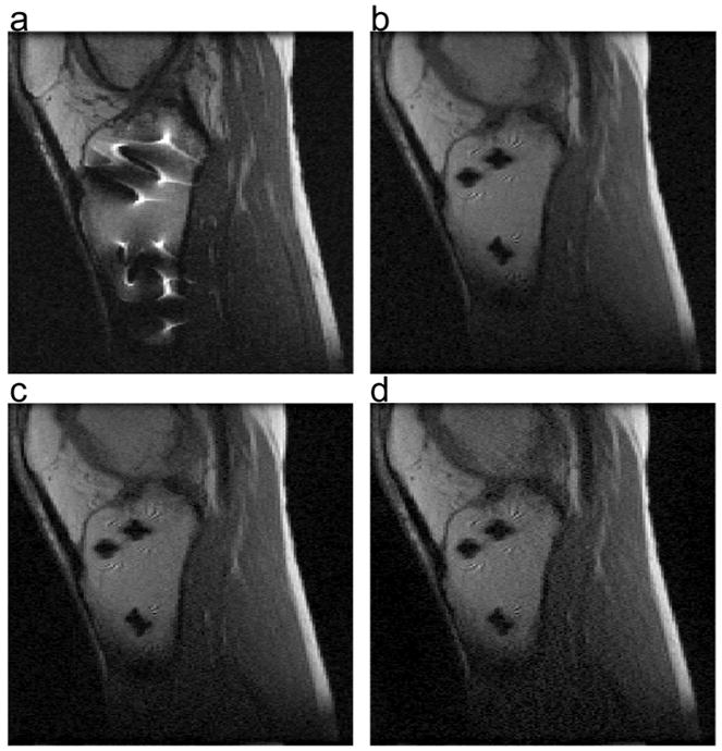
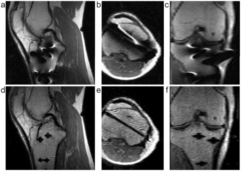
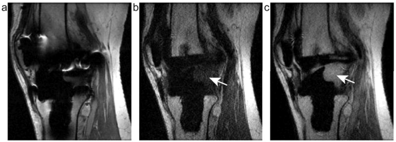
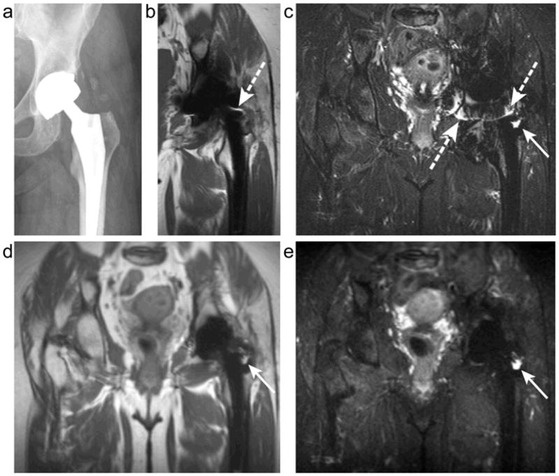
Materials and methods: Slice-encoding for metal artifact correction (SEMAC) is a modified spin echo sequence that uses view-angle tilting and slice-direction phase encoding to correct both in-plane and through-plane artifacts. Standard spin echo trains and short-TI inversion recovery (STIR) allow efficient PD-weighted imaging with optional fat suppression. A completely linear reconstruction allows incorporation of parallel imaging and partial Fourier imaging. The signal-to-noise ratio (SNR) effects of all reconstructions were quantified in one subject. Ten subjects with different metallic implants were scanned using SEMAC protocols, all with scan times below 11 minutes, as well as with standard spin echo methods.

Results: The SNR using standard acceleration techniques is unaffected by the linear SEMAC reconstruction. In all cases with implants, accelerated SEMAC significantly reduced artifacts compared with standard imaging techniques, with no additional artifacts from acceleration techniques. The use of different contrast mechanisms allowed differentiation of fluid from other structures in several subjects.

Conclusion: SEMAC imaging can be combined with standard echo-train imaging, parallel imaging, partial-Fourier imaging, and inversion recovery techniques to offer flexible image contrast with a dramatic reduction of metal-induced artifacts in scan times under 11 minutes.


Figures

Figure 1
Figure 1
SEMAC acquisition and reconstruction. (a) The desired volume and number of sections are prescribed. (b) An arbitrary number of excited slices cover the prescribed volume. For each slice (example highlighted), the phase-encoded FOVz includes enough sections to cover the maximum expected range of slice distortions. (c) The reconstruction Fourier transforms data, to obtain the centered FOVz shown, then uses modulo arithmetic to obtain sections shown covering the FOVz around the excited slice. (d) Simply adding the correctly placed sections to a distortion corrected volume combines the signal from all excited slices. Since the combination is linear, it can precede subsequent in-plane reconstruction steps.
Figure 2
Figure 2
SEMAC pulse sequence. Standard RF waveforms are used, with centered phase-ramps to shift the slice location without altering relative slice-to-slice phase. A VAT gradient is played during the readout to correct in-plane distortions, and SEMAC phase encoding is added to resolve the through-plane location. Multiple CPMG echoes can be used as in standard RARE imaging.
Figure 3
Figure 3
Fully sampled SEMAC image of the neck of a shoulder prosthesis (a), and images comparing the highlighted region with different techniques (b–e) and reformatted images in an orthogonal plane (f–i). Fully sampled SEMAC, 16:26 (b,f), 2× ARC accelerated SEMAC, 10:05 (c,g), and 2× ARC with 60% ky acquisition, 7:22 (d,h). For comparison, a standard spin echo image with the same parameters, 1:06 (e,i), shows substantially more distortion. SEMAC images all perform similarly in terms of distortion correction, showing the neck detail and curved head (arrows).
Figure 4
Figure 4
Measured SNR using SEMAC (red) and standard 2D multi-slice (blue) acquisitions and reconstructions, for 1×, 2× and 3× outer k-space sub-sampling, and with full and 60% ky acquisitions. Hollow boxes show the SNRs normalized so that the 1× scans have equivalent SNR. Solid horizontal bars show the expected normalized SNR due to scan time reduction alone; additional losses are due to “g-factor” noise amplification in parallel imaging, and are much more significant with 3× acceleration than 2× acceleration.
Figure 5
Figure 5
Spin echo and SEMAC images in a volunteer with stainless steel screws below the knee. (a) Standard SE acquisition (2:20) (b) Full SEMAC acquisition (18:48). (c) 2× ARC accelerated SEMAC (11:06). (d) 3× ARC accelerated SEMAC (8:29). Accelerated reconstructions are achieved from a single dataset by discarding data. Noise increases, but scan time drops significantly without degrading the artifact correction.
Figure 6
Figure 6
PD-weighted Knee Images with ARC and a higher matrix: 256 × 128, 32 slices, full k-space 9-minute SEMAC scan with 2× ARC acceleration (a–c) PD-weighted Spin Echo, (d–f) PD-weighted SEMAC. Images are acquired in the sagittal plane (a,d) with axial (b,e) and coronal (c,f) reformats shown. The dramatically improved depiction of metal screws in SEMAC images compared to spin echo images is clear, especially in the axial reformat.
Figure 7
Figure 7
Sagittal images in a volunteer with a total knee arthroplasty comparing PD-weighted FSE to SEMAC with 2×ARC acceleration and 60% partial Fourier acquisition. (a) PD-weighted FSE, 2.5 min, (b) T1-weighted SEMAC, 12 min and (c) PD-weighted SEMAC, 10 min. SEMAC images show that both PD- and T1-weighted contrast can be achieved with depiction of the bone-implant interface. The different contrast shows fluid posterior to the implant (arrows).
Figure 8
Figure 8
Plain film X-ray (a), conventional T1 FSE (b), conventional STIR (c), proton-density SEMAC (d) and STIR SEMAC (e) images in a subject with a total hip replacment. Susceptibility artifacts cause “pile-up” artifacts (dashed arrows) and inaccurate depiction of the implant on conventional MR images. SEMAC images dramatically reduce the distortion, and SEMAC STIR allows fluid near the implant to be unambiguously identified where it appears similar to the metal artifact on conventional images.

Similar articles

Cited by

References

    1. Schenck JF. The role of magnetic susceptibility in magnetic resonance imaging: MRI magnetic compatibility of the first and second kinds. Med Phys. 1996;23:815–850. - PubMed
    1. Cho ZH, Ahn CB, Kim JH, Lee YE, Mun CW. Proc., SMRM, 6th Annual Meeting. New York: 1987. Phase error corrected interlaced echo planar imaging; p. 912.
    1. Chang H, Fitzpatrick J. A technique for accurate magnetic resonance imaging in the presence of field inhomogeneities. IEEE Transactions on medical imaging. 1992;11:319–329. - PubMed
    1. Hoff M, Xiang Q. Proceedings of the 17th Annual Meeting of ISMRM. Honolulu: 2009. Eliminating metal artifact distortion using 3d-place; p. 570.
    1. Olsen RV, Munk PL, Lee MJ, et al. Metal artifact reduction sequence: early clinical applications. Radiographics. 2000;20:699–712. - PubMed

Publication types