

Mini drug pump for ophthalmic use
- PMID: 20373877
- PMCID: PMC2888264
- DOI: 10.3109/02713680903521936
Mini drug pump for ophthalmic use
Abstract
Purpose: To evaluate the feasibility of developing a novel mini drug pump for ophthalmic use.
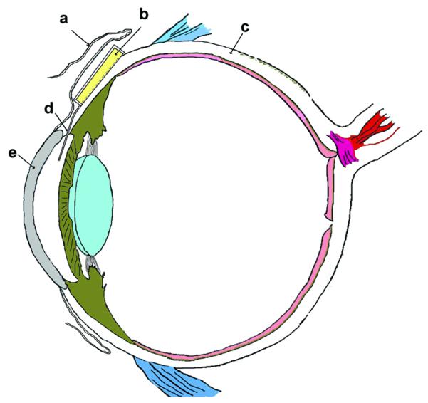
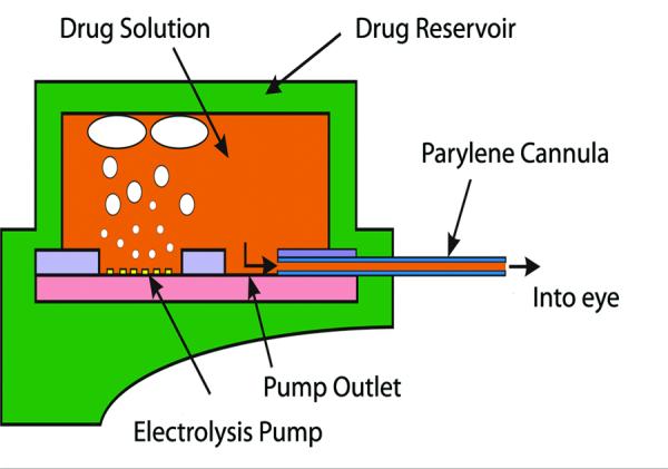
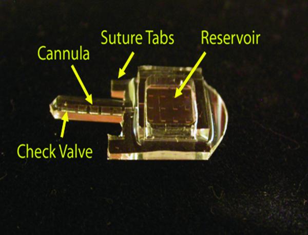
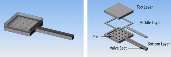
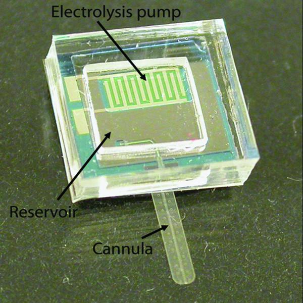
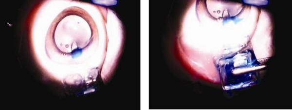
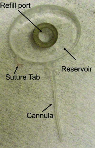
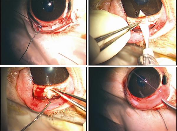
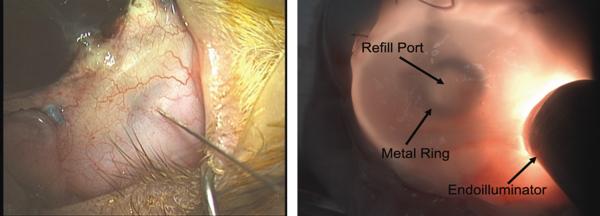
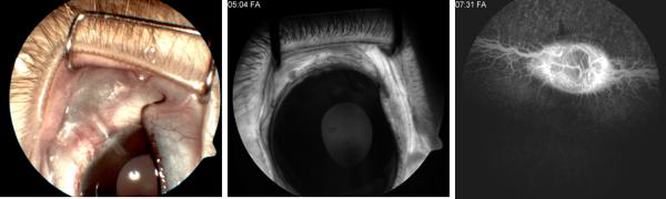
Methods: Using principles of microelectromechanical systems engineering, a mini drug pump was fabricated. The pumping mechanism is based on electrolysis and the pump includes a drug refill port as well as a check valve to control drug delivery. Drug pumps were tested first on the bench-top and then after implantation in rabbits. For the latter, we implanted 4 elliptical (9.9 x 7.7 x 1.8 mm) non-electrically active pumps into 4 rabbits. The procedure is similar to implantation of a glaucoma aqueous drainage device. To determine the ability to refill and also the patency of the cannula, at intervals of 4-6 weeks after implantation, we accessed the drug reservoir with a transconjunctival needle and delivered approximately as low as 1 microL of trypan blue solution (0.06%) into the anterior chamber. Animals were followed by slit lamp examination, photography, and fluorescein angiography.
Results: Bench-top testing showed 2.0 microL/min delivery when using 0.4 mW of power for electrolysis. One-way valves showed reliable opening pressures of 470 mmHg. All implanted devices refilled at 4-6 weeks intervals for 4-6 months. No infection was seen. No devices extruded. No filtering bleb formed over the implant.
Conclusions: A prototype ocular mini drug pump was built, implanted, and refilled. Such a platform needs more testing to determine the long term biocompatibility of an electrically-controlled implanted pump. Testing with various pharmacological agents is needed to determine its ultimate potential for ophthalmic use.
Figures
References
-
- Geroski DH, Edelhauser HF. Drug delivery for posterior segment eye disease. Invest Ophthalmol Vis Sci. 2000;41:961–964. - PubMed
-
- Hoyng PF, van Beek LM. Pharmacological therapy for glaucoma: a review. Drugs. 2000;59:411–434. - PubMed
-
- Hsu J. Drug delivery methods for posterior segment disease. Curr Opin Ophthalmol. 2007;18:235–239. - PubMed
-
- Shell JW. Ophthalmic drug delivery systems. Surv Ophthalmol. 1984;29:117–128. - PubMed
-
- Davies NM. Biopharmaceutical considerations in topical ocular drug delivery. Clin Exp Pharmacol Physiol. 2000;27:558–562. - PubMed
Publication types
MeSH terms
Substances
Grants and funding
LinkOut - more resources
Full Text Sources
Other Literature Sources