Review
. 2010 Apr;17(3):164-78.
doi: 10.1111/j.1549-8719.2010.00025.x.

The effects of hemodynamic force on embryonic development

Affiliations
Review

The effects of hemodynamic force on embryonic development

James C Culver et al. Microcirculation. 2010 Apr.

Abstract

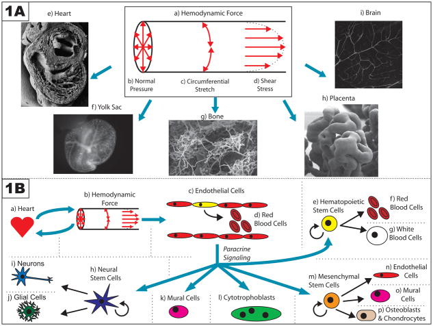
Blood vessels have long been known to respond to hemodynamic force, and several mechanotransduction pathways have been identified. However, only recently have we begun to understand the effects of hemodynamic force on embryonic development. In this review, we will discuss specific examples illustrating the role of hemodynamic force during the development of the embryo, with particular focus on the development of the vascular system and the morphogenesis of the heart. We will also discuss the important functions served by mechanotransduction and hemodynamic force during placentation, as well as in regulating the maintenance and division of embryonic, hematopoietic, neural, and mesenchymal stem cells. Pathological misregulation of mechanosensitive pathways during pregnancy and embryonic development may contribute to the occurrence of cardiovascular birth defects, as well as to a variety of other diseases, including preeclampsia. Thus, there is a need for future studies focusing on better understanding the physiological effects of hemodynamic force during embryonic development and their role in the pathogenesis of disease.


Figures

Figure 1
Figure 1. Summary of the effects of hemodynamic force on embryonic development
(A) Three mutually perpendicular hemodynamic forces are exerted onto the vessel wall by flowing blood (a). First, blood pressure exerts a normal force against the vessel wall (b). Second, as a result of this normal force, the cells comprising the wall also experience circumferential stretch (c). Finally, because flowing blood exerts drag on the vessel wall, the endothelial cells that line the vessel experience a frictional force known as shear stress (d). These hemodynamic forces are important at a variety of developmental time points and in a diverse range of vessel architectures, including those seen in: the heart (e – adapted with permission from Oxford University Press: <Hogers B, et al. (1999). Extraembryonic venous obstructions lead to cardiovascular malformations and can be embryolethal. Cardiovasc Res 41:94. Figure 6a.>) [45]; the extraembryonic yolk sac (f); the bone (g – adapted with kind permission from Springer Science+Business Media: <Morini S, et al. (2006). Microvascular adaptation to growth in rat humeral head. Anat Embryol (Berl) 211:407. Figure 8.>) [82]; the placenta (h – © Society for Reproduction and Fertility (2009). Reproduced by permission. Adapted from: <Burton GJ, et al. (2009). Regulation of vascular growth and function in the human placenta. Reproduction 138:897. Figure 3b.>) [11]; and the brain (i). (B) Hemodynamic force can have effects on a variety of cell types. The heart (a) creates hemodynamic force (b) by pumping blood; reciprocally, this force affects the morphogenesis of the heart as it develops. This hemodynamic force is also sensed by endothelial cells that line the walls of vessels (c), and thereby directs a variety of intrinsic cellular responses, including those that are important for vascular remodeling and arterial/venous specification. Some of these endothelial cells, particularly in the aorta-gonads-mesonephros, may be hemogenic endothelial cells (depicted in yellow) that divide to produce blood cells in a flow-dependent manner (d). Hemodynamic force may also further regulate the self-renewal and differentiation of hematopoietic stem cells that are not part of the vessel wall (e) by using paracrine signals released by the endothelium. Other paracrine signals released by the endothelium in response to hemodynamic force may also influence the self-renewal and differentiation of progenitor cells in other stem cell niches, including neural stem cells (h) and mesenchymal stem cells (m). Finally, flow-dependent release of paracrine signals by endothelial cells have also been implicated in the recruitment of mural cells (k) to the walls of developing vessels, and in the invasion of maternal vessels in the endometrium by cytotrophoblasts (l) during placentation.
Figure 2
Figure 2. Defective yolk sac vascular remodeling in embryos with deficient heart function
Shown are littermate mouse embryos at the 7 somite (A, B), 10 somite (C, D), and 23 somite (E, F) stages, which are either heterozygous (A, C, E) or homozygous (B, D, F) for the recessive null allele of Mlc2a. Blood vessels are visualized through use of the fluorescent reporter Tg(ε-globin-KGFP), which is transgenically expressed in primitive erythroblasts. Shortly after gastrulation, extraembryonic mesodermal cells in the yolk sac coalesce to form the blood islands (A). Shortly thereafter, angioblasts in these blood islands form the primitive capillary plexus of the yolk sac (C), and by the 23 somite stage, this early capillary plexus has remodeled into a hierarchical structure of large and small vessels (E). Mice which are homozygous for the Mlc2a null mutation, and therefore have deficient heart function, are still able to form the blood islands (B) and the primitive capillary plexus in the yolk sac (D). However, they are not able to remodel their yolk sac vasculature as the embryo grows (F), and therefore die in mid-gestation. This process of vascular remodeling is thought to depend on the hemodynamic force created by the robust flow of blood through the capillary plexus in wildtype mice (adapted with permission from the Company of Biologists: <Lucitti JL, et al. (2007). Vascular remodeling of the mouse yolk sac requires hemodynamic force. Development 134:3321. DOI:10.1242/dev.02883. Figure 4.>) [68].
Figure 3
Figure 3. The adult neural stem cell niche
This figure shows a flatmount preparation of the highly vascular subependymal zone of the adult mouse brain. Functional vessels are visualized with a fluorescently labeled dextran that fills the vessel lumen. Neural stem cells persist in this region of the adult brain, which is directly adjacent to the lateral ventricle.

Similar articles

Cited by

References

    1. Adamo L, Naveiras O, Wenzel PL, McKinney-Freeman S, Mack PJ, Gracia-Sancho J, Suchy-Dicey A, Yoshimoto M, Lensch MW, Yoder MC, García-Cardeña G, Daley GQ. Biomechanical forces promote embryonic haematopoiesis. Nature. 2009;459:1131–1135. - PMC - PubMed
    1. Ai D, Liu W, Ma L, Dong F, Lu MF, Wang D, Verzi MP, Cai C, Gage PJ, Evans S, Black BL, Brown NA, Martin JF. Pitx2 regulates cardiac left-right asymmetry by patterning second cardiac lineage-derived myocardium. Dev Biol. 2006;296:437–449. - PMC - PubMed
    1. Aikawa R, Nagai T, Kudoh S, Zou Y, Tanaka M, Tamura M, Akazawa H, Takano H, Nagai R, Komuro I. Integrins play a critical role in mechanical stress-induced p38 MAPK activation. Hypertension. 2002;39:233–238. - PubMed
    1. Armulik A, Abramsson A, Betsholtz C. Endothelial/pericyte interactions. Circ Res. 2005;97:512–523. - PubMed
    1. Arvidsson A, Collin T, Kirik D, Kokaia Z, Lindvall O. Neuronal replacement from endogenous precursors in the adult brain after stroke. Nat Med. 2002;8:963–970. - PubMed

Publication types

MeSH terms