

Mechanobiology and the microcirculation: cellular, nuclear and fluid mechanics
- PMID: 20374482
- PMCID: PMC2881226
- DOI: 10.1111/j.1549-8719.2009.00016.x
Mechanobiology and the microcirculation: cellular, nuclear and fluid mechanics
Abstract
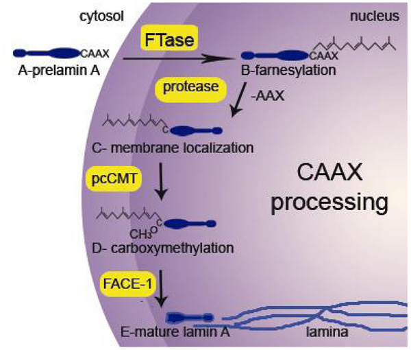
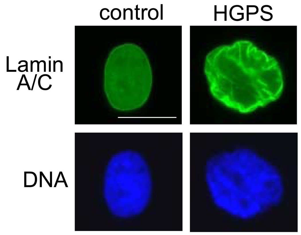
Endothelial cells are stimulated by shear stress throughout the vasculature and respond with changes in gene expression and by morphological reorganization. Mechanical sensors of the cell are varied and include cell surface sensors that activate intracellular chemical signaling pathways. Here, possible mechanical sensors of the cell including reorganization of the cytoskeleton and the nucleus are discussed in relation to shear flow. A mutation in the nuclear structural protein lamin A, related to Hutchinson-Gilford progeria syndrome, is reviewed specifically as the mutation results in altered nuclear structure and stiffer nuclei; animal models also suggest significantly altered vascular structure. Nuclear and cellular deformation of endothelial cells in response to shear stress provides partial understanding of possible mechanical regulation in the microcirculation. Increasing sophistication of fluid flow simulations inside the vessel is also an emerging area relevant to the microcirculation as visualization in situ is difficult. This integrated approach to study--including medicine, molecular and cell biology, biophysics and engineering--provides a unique understanding of multi-scale interactions in the microcirculation.
Figures
Similar articles
-
Mechanics in human fibroblasts and progeria: Lamin A mutation E145K results in stiffening of nuclei.J Mol Recognit. 2017 Feb;30(2). doi: 10.1002/jmr.2580. Epub 2016 Sep 28. J Mol Recognit. 2017. PMID: 27677907
-
Endothelial Nuclear Lamina in Mechanotransduction Under Shear Stress.Adv Exp Med Biol. 2018;1097:83-104. doi: 10.1007/978-3-319-96445-4_5. Adv Exp Med Biol. 2018. PMID: 30315541 Review.
-
Vascular mechanobiology: endothelial cell responses to fluid shear stress.Circ J. 2009 Nov;73(11):1983-92. doi: 10.1253/circj.cj-09-0583. Epub 2009 Oct 5. Circ J. 2009. PMID: 19801852 Review.
-
The nuclear membrane and mechanotransduction: impaired nuclear mechanics and mechanotransduction in lamin A/C deficient cells.Novartis Found Symp. 2005;264:264-73; discussion 273-8. Novartis Found Symp. 2005. PMID: 15773759 Review.
-
Progerin-expressing endothelial cells are unable to adapt to shear stress.Biophys J. 2022 Feb 15;121(4):620-628. doi: 10.1016/j.bpj.2022.01.004. Epub 2022 Jan 6. Biophys J. 2022. PMID: 34999130 Free PMC article.
Cited by
-
Lamins at a glance.J Cell Sci. 2012 May 1;125(Pt 9):2087-93. doi: 10.1242/jcs.087288. J Cell Sci. 2012. PMID: 22669459 Free PMC article. Review. No abstract available.
-
Sustained Physiological Stretch Induces Abdominal Skin Growth in Pregnancy.Ann Biomed Eng. 2024 Jun;52(6):1576-1590. doi: 10.1007/s10439-024-03472-6. Epub 2024 Feb 29. Ann Biomed Eng. 2024. PMID: 38424309 Free PMC article.
-
Progeria: a paradigm for translational medicine.Cell. 2014 Jan 30;156(3):400-7. doi: 10.1016/j.cell.2013.12.028. Cell. 2014. PMID: 24485450 Free PMC article.
-
Recapitulation of molecular regulators of nuclear motion during cell migration.Cell Adh Migr. 2019 Dec;13(1):50-62. doi: 10.1080/19336918.2018.1506654. Epub 2018 Sep 27. Cell Adh Migr. 2019. PMID: 30261154 Free PMC article. Review.
-
Cellular and Molecular Bioengineering: A Tipping Point.Cell Mol Bioeng. 2012 Sep 1;5(3):239-253. doi: 10.1007/s12195-012-0246-7. Epub 2012 Aug 24. Cell Mol Bioeng. 2012. PMID: 23264805 Free PMC article.
References
-
- Adelfalk C, Scherthan H, Hirsch-Kauffmann M, Schweiger M. Nuclear deformation characterizes Werner syndrome cells. Cell Biol Int. 2005;29:1032–1037. - PubMed
-
- AlMomani T, Udaykumar HS, Marshall JS, Chandran KB. Micro-scale dynamic simulation of erythrocyte-platelet interaction in blood flow. Ann Biomed Eng. 2008;36:905–920. - PubMed
-
- Bagchi P, Johnson PC, Popel AS. Computational fluid dynamic simulation of aggregation of deformable cells in a shear flow. J Biomech Eng. 2005;127:1070–1080. - PubMed
-
- Barakat AI, Leaver EV, Pappone PA, Davies PF. A flow-activated chloride-selective membrane current in vascular endothelial cells. Circ Res. 1999;85:820–828. - PubMed
-
- Barbee JH, Cokelet GR. Prediction of blood flow in tubes with diameters as small as 29 microns. Microvasc Res. 1971;3:17–21. - PubMed
Publication types
MeSH terms
Substances
Grants and funding
LinkOut - more resources
Full Text Sources
