Tumor microvasculature and microenvironment: novel insights through intravital imaging in pre-clinical models
- PMID: 20374484
- PMCID: PMC2859831
- DOI: 10.1111/j.1549-8719.2010.00029.x
Tumor microvasculature and microenvironment: novel insights through intravital imaging in pre-clinical models
Abstract
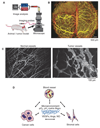
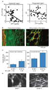
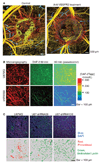
Intravital imaging techniques have provided unprecedented insight into tumor microcirculation and microenvironment. For example, these techniques allowed quantitative evaluations of tumor blood vasculature to uncover its abnormal organization, structure and function (e.g., hyper-permeability, heterogeneous and compromised blood flow). Similarly, imaging of functional lymphatics has documented their absence inside tumors. These abnormalities result in elevated interstitial fluid pressure and hinder the delivery of therapeutic agents to tumors. In addition, they induce a hostile microenvironment characterized by hypoxia and acidosis, as documented by intravital imaging. The abnormal microenvironment further lowers the effectiveness of anti-tumor treatments such as radiation therapy and chemotherapy. In addition to these mechanistic insights, intravital imaging may also offer new opportunities to improve therapy. For example, tumor angiogenesis results in immature, dysfunctional vessels--primarily caused by an imbalance in production of pro- and anti-angiogenic factors by the tumors. Restoring the balance of pro- and anti-angiogenic signaling in tumors can "normalize" tumor vasculature and thus, improve its function, as demonstrated by intravital imaging studies in preclinical models and in cancer patients. Administration of cytotoxic therapy during periods of vascular normalization has the potential to enhance treatment efficacy.
Figures


References
-
- Alexandrakis G, Brown EB, Tong RT, et al. Two-photon fluorescence correlation microscopy reveals the two-phase nature of transport in tumors. Nat Med. 2004;10:203–207. - PubMed
-
- Baish JW, Jain RK. Fractals and cancer. Cancer Res. 2000;60:3683–3688. - PubMed
-
- Berk DA, Swartz MA, Leu AJ, Jain RK. Transport in lymphatic capillaries. II. Microscopic velocity measurement with fluorescence photobleaching. Am J Physiol. 1996;270:H330–H337. - PubMed
Publication types
MeSH terms
Substances
Grants and funding
- R01 CA126642/CA/NCI NIH HHS/United States
- R01-CA085140/CA/NCI NIH HHS/United States
- R01 CA149285/CA/NCI NIH HHS/United States
- R01 CA085140/CA/NCI NIH HHS/United States
- P01-CA-080124/CA/NCI NIH HHS/United States
- R21 CA139168/CA/NCI NIH HHS/United States
- R01-CA115767/CA/NCI NIH HHS/United States
- R24 CA085140/CA/NCI NIH HHS/United States
- R01-CA096915/CA/NCI NIH HHS/United States
- R13 CA119852/CA/NCI NIH HHS/United States
- R01 CA115767/CA/NCI NIH HHS/United States
- R01 CA096915/CA/NCI NIH HHS/United States
- P01 CA080124/CA/NCI NIH HHS/United States
- R01-CA126642/CA/NCI NIH HHS/United States
- R01 CA159258/CA/NCI NIH HHS/United States
- R21-139168/PHS HHS/United States
LinkOut - more resources
Full Text Sources
Other Literature Sources
Medical

