. 2010 Apr 7:8:33.
doi: 10.1186/1741-7007-8-33.

The lethal giant larvae tumour suppressor mutation requires dMyc oncoprotein to promote clonal malignancy

Affiliations

The lethal giant larvae tumour suppressor mutation requires dMyc oncoprotein to promote clonal malignancy

Francesca Froldi et al. BMC Biol. .

Abstract

Background: Neoplastic overgrowth depends on the cooperation of several mutations ultimately leading to major rearrangements in cellular behaviour. Precancerous cells are often removed by cell death from normal tissues in the early steps of the tumourigenic process, but the molecules responsible for such a fundamental safeguard process remain in part elusive. With the aim to investigate the molecular crosstalk occurring between precancerous and normal cells in vivo, we took advantage of the clonal analysis methods that are available in Drosophila for studying the phenotypes due to lethal giant larvae (lgl) neoplastic mutation induced in different backgrounds and tissues.

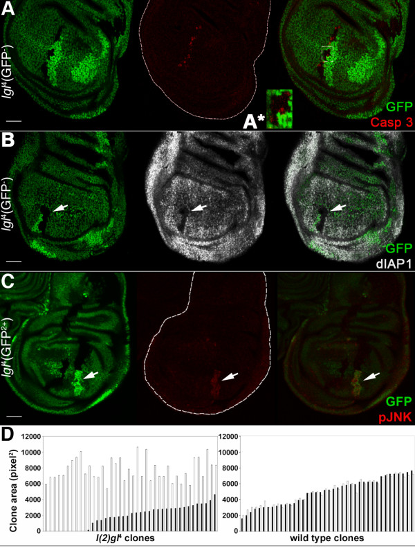
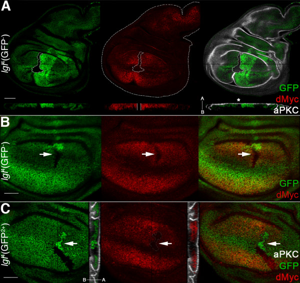
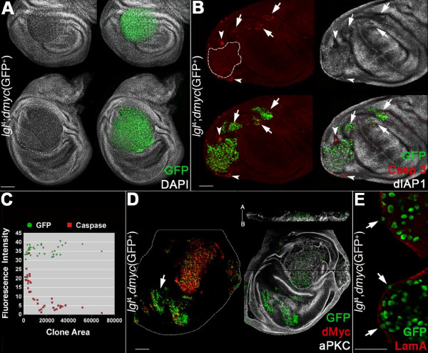
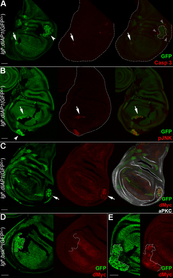
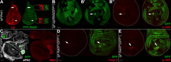
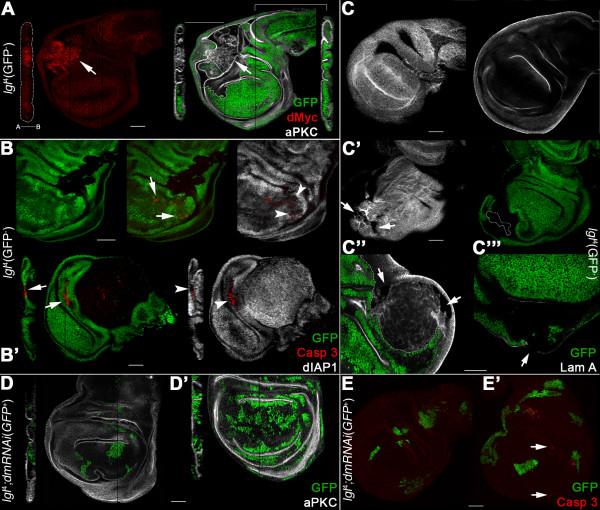
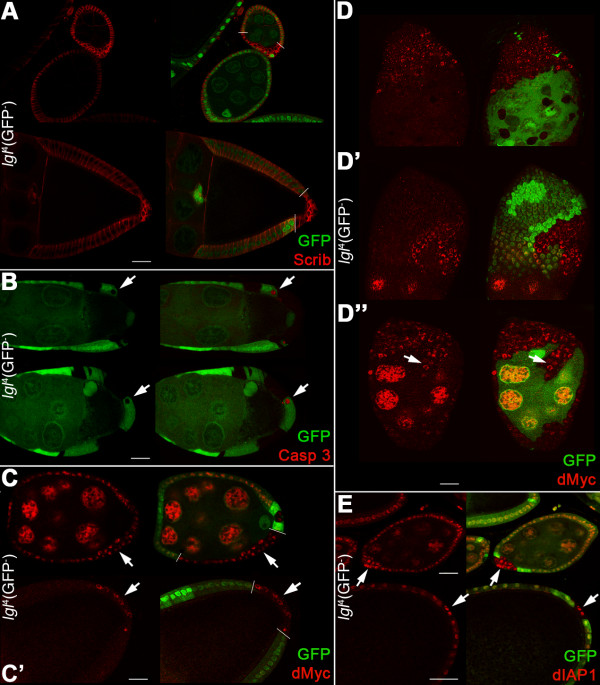
Results: We observed that lgl mutant cells growing in wild-type imaginal wing discs show poor viability and are eliminated by Jun N-terminal Kinase (JNK)-dependent cell death. Furthermore, they express very low levels of dMyc oncoprotein compared with those found in the surrounding normal tissue. Evidence that this is a cause of lgl mutant cells elimination was obtained by increasing dMyc levels in lgl mutant clones: their overgrowth potential was indeed re-established, with mutant cells overwhelming the neighbouring tissue and forming tumourous masses displaying several cancer hallmarks. Moreover, when lgl mutant clones were induced in backgrounds of slow-dividing cells, they upregulated dMyc, lost apical-basal cell polarity and were able to overgrow. Those phenotypes were abolished by reducing dMyc levels in the mutant clones, thereby confirming its key role in lgl-induced tumourigenesis. Furthermore, we show that the eiger-dependent Intrinsic Tumour Suppressor pathway plays only a minor role in eliminating lgl mutant cells in the wing pouch; lgl-/- clonal death in this region is instead driven mainly by dMyc-induced Cell Competition.

Conclusions: Our results provide the first evidence that dMyc oncoprotein is required in lgl tumour suppressor mutant tissue to promote invasive overgrowth in larval and adult epithelial tissues. Moreover, we show that dMyc abundance inside versus outside the mutant clones plays a key role in driving neoplastic overgrowth.


Figures

Figure 1
Figure 1
lgl-/- clones induced in lgl+/- imaginal wing discs die by apoptosis and the surrounding tissue grows at their expense. A, B: lgl-/- clones (GFP-) induced in a w, hs-Flp/+; l(2)gl4, FRT40A/Ubi>GFPnls, FRT40A background (GFP+). Wild-type twin clones are GFP2+. A: active-Caspase 3 staining; in A*, a magnification of the region outlined is shown. B: dIAP1 staining; its expression within the mutant clone (arrow) is visibly lower. C: lgl-/- clones (GFP2+) induced in a w, hs-Flp/+; l(2)gl4, Ubi>GFPnls, FRT40A/FRT40A background (GFP+). Wild-type twin clones are GFP-. pJNK staining shows that the JNK pathway is activated inside the lgl mutant clone (arrow). Wing discs are outlined in A and C. Scale bars are 35 μm. Mutant clone genotypes are indicated. D: lgl-/- (left panel) and wild-type (right panel) clone profile from a twin analysis of l(2)gl4 or wild-type clones sampled in the wing pouch region induced in a w, hs-Flp/+; l(2)gl4, FRT40A or FRT40A/Ubi>GFPnls, FRT40A background. Black bars indicate lgl4 (n = 40) and wild-type (n = 40) clones and white bars indicate the respective twins. For this experiment, freshly hatched larvae were collected in a one-hour time window and staged on cornmeal medium to 90 hours after hatching before collecting tissues.
Figure 2
Figure 2
lgl mutant clones show low levels of dMyc oncoprotein with respect to the surrounding tissue. A, B: lgl-/- clones (GFP-) induced in a w, hs-Flp/+; l(2)gl4, FRT40A/Ubi>GFPnls, FRT40A background (GFP+). Wild-type twin clones are GFP2+. A: dMyc and aPKC staining; clone is outlined and the projection along the Z axis shows that dMyc expression within the mutant clone is low all along the disc thickness (enclosed between two white bars) and does not show strong defects in apical-basal cell polarity (asterisk). The apical-basal axis of the disc proper is also shown. Another lgl-/- clone showing low dMyc levels (arrow) can be observed in B at higher magnification. C: To show mutant nuclei, lgl-/- clones (GFP2+, arrow) were also induced in a w, hs-Flp/+; l(2)gl4, Ubi>GFPnls, FRT40A/FRT40A background (GFP+). Wild-type twin clones are GFP-. In the projection along the Z axis it can be seen that lgl-/- cells are being basally extruded from the epithelium. Wing disc is outlined in A. Scale bars are 35 μm. Mutant clone genotypes are indicated.
Figure 3
Figure 3
dMyc expression in lgl-/- clones induced in an lgl+/- background promotes overgrowth and invasive behaviour. A-E: lgl-/-; UAS-dmyc clones (GFP+) in a yw, hs-Flp, tub>Gal4, UAS-GFP/+; l(2)gl4, FRT40A/tub>Gal80, FRT40A; UAS-dmyc/+ background (GFP-). A: clone morphology. B: active-Caspase 3 and dIAP1 staining; the arrows indicate groups of mutant cells undergoing autonomous apoptosis and the arrowheads indicate lgl+/- cells dying outside a mutant clone. C: distribution of the ratios 'clone area/active Caspase 3 fluorescence intensity' (red squares) and 'clone area/GFP fluorescence intensity' (green circles) as a control (n = 40); D: dMyc and aPKC staining; the projection along the Z axis shows the multilayered nature of the epithelium inside the mutant clone. The apical-basal axis of the disc proper is also indicated. The arrows indicate clones in the wing pouch that show low levels of dMyc protein. E: Laminin A staining; the arrows indicate regions of discontinuity. Wing disc is outlined in D. Scale bars are 35 μm. Mutant clone genotypes are indicated.
Figure 4
Figure 4
Inhibition of cell death is not sufficient to unveil the malignant potential of the lgl-/- clones in the wing pouch region. A-C: lgl-/-; UAS-dIAP clones (GFP2+) induced in a yw, hs-Flp, tub>Gal4/+; l(2)gl4, Ubi>GFPnls, FRT40A/tub>Gal80, FRT40A; UAS-dIAP/+ background (GFP+). Wild-type twin clones are GFP-. Active-Caspase 3 (A) and pJNK (B) signals are both visible inside lgl mutant clones (arrows). Arrowheads indicate clones in the proximal regions and grey arrowheads point to active-Caspase 3 signals in lgl+/- cells surrounding the mutant clone (outlined). In C, an lgl-/- clone expressing high levels of dMyc protein is shown (arrow). D, E: UAS-bskDN; lgl-/- clones (GFP2+) induced in a yw, hs-Flp, tub>Gal4/UAS-bskDN; l(2)gl4, Ubi>GFPnls, FRT40A/tub>Gal80, FRT40 background (GFP+). Wild-type twin clones are GFP-. dMyc protein is low in the mutant clone in the wing pouch (D) but is high in the clone in the pleura (E). Clone boundaries are indicated by the white dotted line. Wing discs are outlined in A and B. Scale bars are 35 μm. Mutant clone genotypes are indicated.
Figure 5
Figure 5
Intrinsic Tumour Suppression is not responsible for lgl-/- clonal death in the wing pouch region. A: Lgl antibody staining (red) of lgl-/- clones (black) induced in a yw, hs-Flp, tub>Gal4/+; l(2)gl4, FRT40A/tub>Gal80, FRT40; tub>YFP::Rab5/+background (red+). The wild-type twin clones are red2+. The arrow indicates an lgl mutant clone in the notum where the YFP::Rab5 signal is slightly increased and the arrowhead indicates an lgl mutant clone in the pouch where the YFP::Rab5 signal is comparable to that found in the surrounding lgl+/- tissue. Magnifications of the mutant clones are shown on the right. B-B": lgl-/-;UAS-egrRNAi clones (GFP2+) induced in a yw, hs-Flp, tub>Gal4/+; l(2)gl4, Ubi>GFPnls, FRT40A/tub>Gal80, FRT40A; UAS-egrRNA i/+ background (GFP+). Wild-type twin clones are GFP-. B and B' show the apical and basal sections of the same disc; in the basal section a mutant clone contains cells that are basally excluded from the epithelium (arrow) and shows pJNK staining (B", arrow). The clone contour is outlined in B. C:, lgl-/-; UAS-YFP::Rab5DN clones (GFP+) induced in a yw, hs-Flp, tub>Gal4, UAS-GFP/+; l(2)gl4, FRT40A/tub>Gal80, FRT40A; UAS-YFP::Rab5DN/+ background (GFP-), stained with aPKC and dMyc. Due to high staining intensity inside the mutant clones, dMyc signal has been kept very low to avoid overexposure, so its endogenous expression is barely visible. D, E: lgl-/-; UAS-YFP::Rab5DN clones (GFP2+) induced in a yw, hs-Flp, tub>Gal4/+; l(2)gl4, Ubi>GFPnls, FRT40A/tub>Gal80, FRT40A; UAS-YFP::Rab5DN/+ background (GFP+). Wild-type twin clones are GFP-. D: active-Caspase 3; E: pJNK. Wing discs are outlined in B", D and E. Scale bars are 35 μm. Mutant clone genotypes are indicated.
Figure 6
Figure 6
lgl-/- malignant behaviour in a Minute background depends on dMyc protein levels. A-C"': lgl-/- clones (GFP-) in a w, hs-Flp/+;l(2)gl4, FRT40A/M(2)24F, Ubi>GFPnls, FRT40A background (GFP+). A: dMyc and aPKC staining. A strong dMyc accumulation is visible in the part of the mutant clone located in the hinge region (arrows). A Z projection of the mutant clone shows that loss of cell polarity accompanies the dramatic overgrowth; the apical-basal axis of the disc proper is shown. A Z projection of a region of the disc without mutant clones is shown on the right as a control. B: Activated Caspase 3 and dIAP1 staining. Arrows indicate M/+, lgl+/- cells dying around the mutant clones; the Z projection confirms that dying cells are outside the lgl-/- tissue. Arrowheads indicate that dying cells are deprived of dIAP1. C: Surface and cross sections of a wild-type disc stained with Laminin A. C'-C"': Laminin A staining of lgl-/- clones in a M/+, lgl+/- background; arrows indicate several points in which basement membrane integrity is lost. D-E': lgl-/-; UAS-dmRNAi clones (GFP+) in a yw, hs-Flp, tub>Gal4, UAS-GFP/+;l(2)gl4, FRT40A/M(2)24F, tub>Gal80, FRT40A; UAS-dmRNAi/+ background (GFP-). D, D': aPKC staining reveals that lgl-/-; UAS-dmRNAi mutant cells in a M/+, lgl+/- background do not lose apical-basal polarity (compare with 6A). Heat shock pulses were 20 minutes (D) and 1 hour duration (D'). E, E': Caspase staining shows that apoptosis is either absent (E) or scattered throughout the disc (E', arrows). Scale bars are 35 μm. Mutant clone genotypes are indicated.
Figure 7
Figure 7
lgl-/- clonal overgrowth is sustained by dMyc expression also in the follicular epithelium. A-E: Twin-clone analysis of lgl-/- clones (GFP-) induced in a w, hs-Flp/+; l(2)gl4, FRT40A/Ubi>GFPnls, FRT40A background (GFP+). Wild-type twin clones are GFP2+. A: lgl-/- clones (bordered by white bars) stained for Scrib are shown in early and late egg chambers. Scrib distribution spreads from lateral to cortical as mutant cells become round. B: active-Caspase 3 staining of Stage 8 to 9 egg chambers; arrows indicate wild-type cells undergoing apoptosis. C: dMyc staining of early (C) and late (C') egg chambers. Bars mark clone boundaries and arrows indicate dMyc expression inside the mutant clones. D-D": dMyc staining in older lgl-/- clones; in D" the arrow indicates mutant cells migrating into the nurse cell territory. E: dIAP1 staining of early and late egg chambers. The arrows indicate high dIAP1 expression in the lgl-/- clones. Mutant clone genotypes are indicated.

Similar articles

Cited by

References

    1. Humbert P, Russel S, Richardson H. Dgl, Scribble and Lgl in cell polarity, cell proliferation and cancer. Bioessays. 2003;25:542–553. doi: 10.1002/bies.10286. - DOI - PubMed
    1. Bilder D. Epithelial polarity and proliferation control: links from the Drosophila neoplastic tumor suppressors. Genes Dev. 2004;18:1909–1925. doi: 10.1101/gad.1211604. - DOI - PubMed
    1. Dow LE, Humbert PO. Polarity regulators and the control of epithelial architecture, cell migration, and tumorigenesis. Int Rev Cytol. 2007;262:253–302. doi: 10.1016/S0074-7696(07)62006-3. - DOI - PubMed
    1. Woodhouse E, Hersperger E, Shearn A. Growth, metastasis and invasiveness of Drosophila tumors caused by mutations in specific tumor suppressor genes. Dev Genes Evol. 1998;207:542–550. doi: 10.1007/s004270050145. - DOI - PubMed
    1. Grifoni D, Garoia F, Schimanski CC, Schmitz G, Laurenti E, Galle PR, Pession A, Cavicchi S, Strand S. The human protein Hugl-1 substitutes for Drosophila lethal giant larvae tumour suppressor function in vivo. Oncogene. 2004;23:8688–8694. doi: 10.1038/sj.onc.1208023. - DOI - PubMed

Publication types

MeSH terms

LinkOut - more resources