Review
. 2010 Apr 7:7:29.
doi: 10.1186/1742-4690-7-29.

HIV-1 assembly in macrophages

Affiliations
Review

HIV-1 assembly in macrophages

Philippe Benaroch et al. Retrovirology. .

Abstract

The molecular mechanisms involved in the assembly of newly synthesized Human Immunodeficiency Virus (HIV) particles are poorly understood. Most of the work on HIV-1 assembly has been performed in T cells in which viral particle budding and assembly take place at the plasma membrane. In contrast, few studies have been performed on macrophages, the other major target of HIV-1. Infected macrophages represent a viral reservoir and probably play a key role in HIV-1 physiopathology. Indeed macrophages retain infectious particles for long periods of time, keeping them protected from anti-viral immune response or drug treatments. Here, we present an overview of what is known about HIV-1 assembly in macrophages as compared to T lymphocytes or cell lines.Early electron microscopy studies suggested that viral assembly takes place at the limiting membrane of an intracellular compartment in macrophages and not at the plasma membrane as in T cells. This was first considered as a late endosomal compartment in which viral budding seems to be similar to the process of vesicle release into multi-vesicular bodies. This view was notably supported by a large body of evidence involving the ESCRT (Endosomal Sorting Complex Required for Transport) machinery in HIV-1 budding, the observation of viral budding profiles in such compartments by immuno-electron microscopy, and the presence of late endosomal markers associated with macrophage-derived virions. However, this model needs to be revisited as recent data indicate that the viral compartment has a neutral pH and can be connected to the plasma membrane via very thin micro-channels. To date, the exact nature and biogenesis of the HIV assembly compartment in macrophages remains elusive. Many cellular proteins potentially involved in the late phases of HIV-1 cycle have been identified; and, recently, the list has grown rapidly with the publication of four independent genome-wide screens. However, their respective roles in infected cells and especially in macrophages remain to be characterized. In summary, the complete process of HIV-1 assembly is still poorly understood and will undoubtedly benefit from the ongoing explosion of new imaging techniques allowing better time-lapse and quantitative studies.


Figures

Figure 1
Figure 1
A current view of HIV assembly in macrophages. The viral genomic RNA transcribed in the nucleus is exported to the cytoplasm. The transmembrane envelope (Env) protein is produced in the endoplasmic reticulum and transits through the Golgi apparatus while Gag is synthesized on free cytosolic ribosomes. Both Env and the Gag precursors are targeted to the assembly site through unidentified pathways. The sites of Gag/Env interaction, Gag multimerization and binding to viral genomic RNA remain elusive as well. The main cellular factors suspected to play a role in these trafficking events are indicated; nevertheless most of the time their roles have still to be established in macrophages. The assembly process requires the hijacking of the cellular ESCRT machinery and occurs on cholesterol- and tetraspanin-enriched membrane microdomains. The assembly compartment can be connected at least transiently to the plasma membrane through thin microchannels that do not allow virion passage. The limiting membrane of the viral assembly compartment as well as the microchannels often exhibit thick molecular coats of which the composition remains obscure. See text for details.
Figure 2
Figure 2
Immunofluorescent staining of Gag in a HIV-1-infected macrophage. Monocyte-derived-macrophages were infected with HIV-1 NLAD8 pseudotyped with VSV-G. At day 7 post-infection, cells were fixed, permeabilized and stained with a rabbit antiserum anti-HIV-1 p17 (AIDS Research and Reference Reagent Program, Division of AIDS, NIAID, NIH, from Dr. Paul Spearman) revealed by goat anti-rabbit antibodies conjugated to Alexa Fluor 488. A three dimensional reconstruction built from an 8 μm thick section (0.5 μm between planes) is presented. It has been generated using the Nikon A1R Confocal laser microscope system. The macrophages often appear with this typical shape in "sunny side up egg" where the nucleus is a small part of the "yolk". The Gag staining appears rich and complex; there is a diffuse cytosolic staining, some structures with intense staining located in the "yolk" which may correspond to the viral assembly compartments, and very small dots scattered everywhere which could correspond to free virions or Gag multimers (the microscope resolution is not good enough to estimate their precise size). Scale bar, 5 μm.
Figure 3
Figure 3
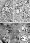
Localization of Alix and CHMP4 at the viral assembly compartment. Monocyte-derived-macrophages infected with HIV-1 NLAD8 for 14 days were processed for cryosectioning as described [65]. (A) Two examples of virus-containing compartments that were triple labeled for p17/p55 Gag with protein A coupled to gold particles of 5 nm or PAG5, for Alix with PAG10, and for CD63 with PAG15. Alix labeling was found on the virions and at the limiting membrane of the viral assembly compartment (black arrowheads). Note the labeled mitochondria nearby (small arrow). (B) Cryosections were triple labeled for p17/p55 Gag with PAG5, for CHMP4B with PAG10, and for CD63 with PAG15. CHMP4B was present in many virions (black arrowheads). In panels (A) and (B), CD63 was at the limiting membrane of the compartment, in small internal vesicles or incorporated in the membrane of virus particles. (B') Two examples of viral compartments double labeled for CHMP4B with PAG10, and p17/p55 Gag with PAG15. CHMP4B was associated with a thick molecular coat present at the limiting membrane of the assembly compartments (black arrowheads). Bars, 100 nm.
Figure 4
Figure 4
Ruthenium red staining of HIV-1 infected macrophages. Monocyte-derived-macrophages infected with HIV-1 (NLAD8) for 14 days were fixed on ice in the presence of ruthenium red (RR) dye and embedded in Epon for transmission electron microscopy as described [65]. (A) Viral assembly compartments negative for the RR dye were observed such as the one which is framed. Electron-dense deposits of ruthenium red-positive material were seen in lipids droplets, which lied deep within macrophages and were especially numerous near HIV-1 virus-containing vacuoles (see white asterisks). However, a majority of virus-containing compartments remained RR negative (see black asterisks). (A') Enlargement of the framed area in A. (B) Viral assembly compartments containing viral particles positive for the RR dye were also observed. Note the presence of a microchannel emanating from the central compartment (black arrowhead). (C) A "sponge-like structure" is shown in the center of the panel exhibiting highly interconnected membranes. Such structures were positive for the RR dye and very frequently were found in the vicinity of viral compartments (see above the structure). Below the structure, note the presence of numerous secondary lysosomes containing small osmiophilic particles (a few examples are pointed by black arrowheads). Bars, 400 nm.

Similar articles

Cited by

References

    1. Gartner S, Markovits P, Markovitz DM, Kaplan MH, Gallo RC, Popovic M. The role of mononuclear phagocytes in HTLV-III/LAV infection. Science. 1986;233:215–219. doi: 10.1126/science.3014648. - DOI - PubMed
    1. Orenstein JM, Meltzer MS, Phipps T, Gendelman HE. Cytoplasmic assembly and accumulation of human immunodeficiency virus types 1 and 2 in recombinant human colony-stimulating factor-1-treated human monocytes: an ultrastructural study. J Virol. 1988;62:2578–2586. - PMC - PubMed
    1. Sharova N, Swingler C, Sharkey M, Stevenson M. Macrophages archive HIV-1 virions for dissemination in trans. Embo J. 2005;24:2481–2489. doi: 10.1038/sj.emboj.7600707. - DOI - PMC - PubMed
    1. Gonzalez-Scarano F, Martin-Garcia J. The neuropathogenesis of AIDS. Nat Rev Immunol. 2005;5:69–81. doi: 10.1038/nri1527. - DOI - PubMed
    1. Ellery PJ, Tippett E, Chiu YL, Paukovics G, Cameron PU, Solomon A, Lewin SR, Gorry PR, Jaworowski A, Greene WC, Sonza S, Crowe SM. The CD16+ monocyte subset is more permissive to infection and preferentially harbors HIV-1 in vivo. J Immunol. 2007;178:6581–6589. - PubMed

Publication types

LinkOut - more resources