

Phospholipase A2 inhibitors protect against prion and Abeta mediated synapse degeneration
- PMID: 20374666
- PMCID: PMC2865460
- DOI: 10.1186/1750-1326-5-13
Phospholipase A2 inhibitors protect against prion and Abeta mediated synapse degeneration
Abstract
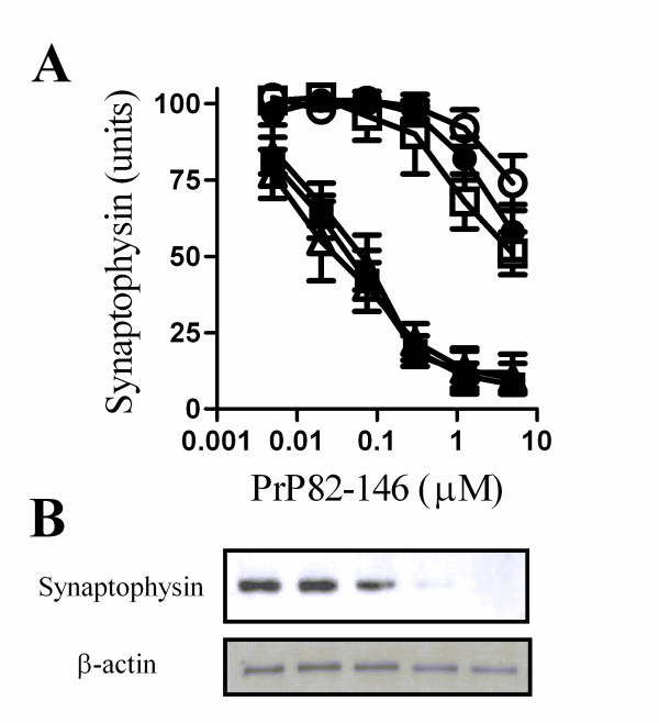
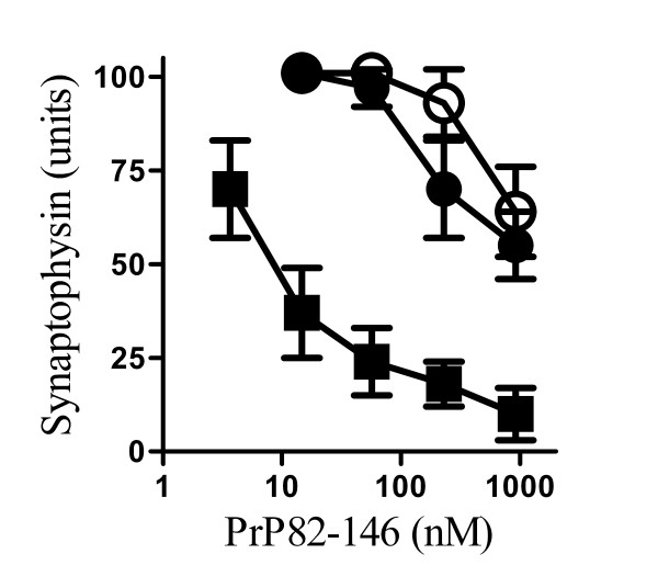
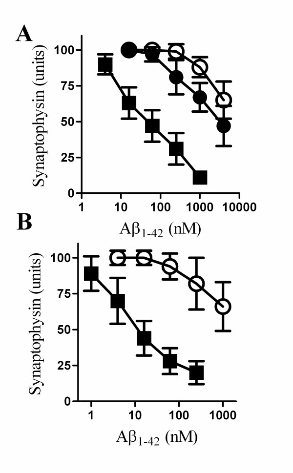
Background: An early event in the neuropathology of prion and Alzheimer's diseases is the loss of synapses and a corresponding reduction in the level of synaptophysin, a pre-synaptic membrane protein essential for neurotransmission. The molecular mechanisms involved in synapse degeneration in these diseases are poorly understood. In this study the process of synapse degeneration was investigated by measuring the synaptophysin content of cultured neurones incubated with the prion derived peptide (PrP82-146) or with Abeta1-42, a peptide thought to trigger pathogenesis in Alzheimer's disease. A pharmacological approach was used to screen cell signalling pathways involved in synapse degeneration.
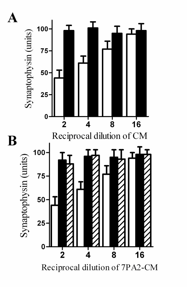
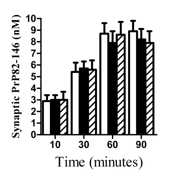
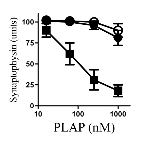
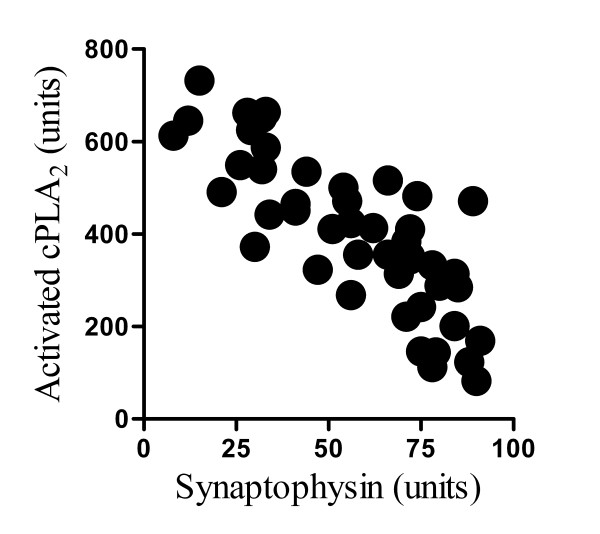
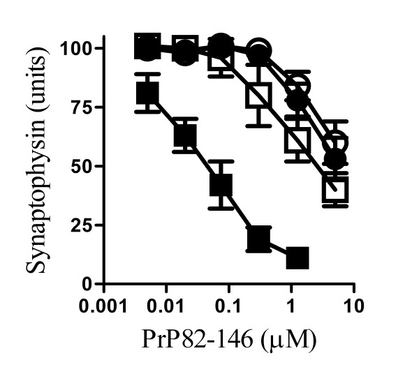
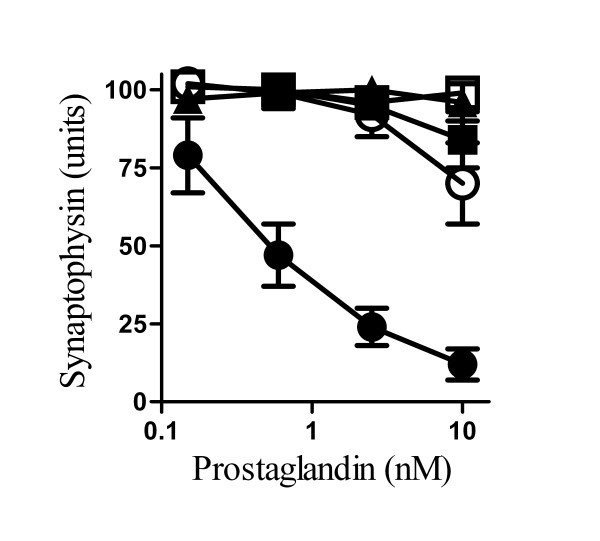
Results: Pre-treatment with phospholipase A2 inhibitors (AACOCF3, MAFP and aristolochic acids) protected against synapse degeneration in cultured cortical and hippocampal neurones incubated with PrP82-146 or Abeta1-42. Synapse degeneration was also observed following the addition of a specific phospholipase A2 activating peptide (PLAP) and the addition of PrP82-146 or Abeta1-42 activated cytoplasmic phospholipase A2 within synapses. Activation of phospholipase A2 is the first step in the generation of platelet-activating factor (PAF) and PAF receptor antagonists (ginkgolide B, Hexa-PAF and CV6029) protected against synapse degeneration induced by PrP82-146, Abeta1-42 and PLAP. PAF facilitated the production of prostaglandin E2, which also caused synapse degeneration and pre-treatment with the prostanoid E receptor antagonist AH13205 protected against PrP82-146, Abeta1-42 and PAF induced synapse degeneration.
Conclusions: Our results are consistent with the hypothesis that PrP82-146 and Abeta1-42trigger abnormal activation of cytoplasmic phospholipase A2 resident within synapses, resulting in elevated levels of PAF and prostaglandin E2that cause synapse degeneration. Inhibitors of this pathway that can cross the blood brain barrier may protect against the synapse degeneration seen during Alzheimer's or prion diseases.
Figures
Similar articles
-
Ginkgolides protect against amyloid-beta1-42-mediated synapse damage in vitro.Mol Neurodegener. 2008 Jan 7;3:1. doi: 10.1186/1750-1326-3-1. Mol Neurodegener. 2008. PMID: 18179689 Free PMC article.
-
Polyunsaturated fatty acids protect against prion-mediated synapse damage in vitro.Neurotox Res. 2010 Apr;17(3):203-14. doi: 10.1007/s12640-009-9093-2. Epub 2009 Jul 31. Neurotox Res. 2010. PMID: 19644728
-
Inhibition of phospholipase A2 increased the removal of the prion derived peptide PrP82-146 from cultured neurons.Neuropharmacology. 2011 Feb-Mar;60(2-3):365-72. doi: 10.1016/j.neuropharm.2010.10.001. Epub 2010 Oct 8. Neuropharmacology. 2011. PMID: 20934441
-
The neuromessenger platelet-activating factor in plasticity and neurodegeneration.Prog Brain Res. 1998;118:281-91. doi: 10.1016/s0079-6123(08)63215-x. Prog Brain Res. 1998. PMID: 9932449 Review.
-
Mediators of injury in neurotrauma: intracellular signal transduction and gene expression.J Neurotrauma. 1995 Oct;12(5):791-814. doi: 10.1089/neu.1995.12.791. J Neurotrauma. 1995. PMID: 8594208 Review.
Cited by
-
Enhanced neuronal degradation of amyloid-β oligomers allows synapse regeneration.Neural Regen Res. 2015 May;10(5):700-1. doi: 10.4103/1673-5374.156955. Neural Regen Res. 2015. PMID: 26109937 Free PMC article. No abstract available.
-
cAMP-Inhibits Cytoplasmic Phospholipase A₂ and Protects Neurons against Amyloid-β-Induced Synapse Damage.Biology (Basel). 2015 Sep 16;4(3):591-606. doi: 10.3390/biology4030591. Biology (Basel). 2015. PMID: 26389963 Free PMC article.
-
Srf1 is a novel regulator of phospholipase D activity and is essential to buffer the toxic effects of C16:0 platelet activating factor.PLoS Genet. 2011 Feb 10;7(2):e1001299. doi: 10.1371/journal.pgen.1001299. PLoS Genet. 2011. PMID: 21347278 Free PMC article.
-
Amyloid-β-induced synapse damage is mediated via cross-linkage of cellular prion proteins.J Biol Chem. 2011 Nov 4;286(44):37955-37963. doi: 10.1074/jbc.M111.248724. Epub 2011 Sep 7. J Biol Chem. 2011. PMID: 21900234 Free PMC article.
-
Breaking the Cycle, Cholesterol Cycling, and Synapse Damage in Response to Amyloid-β.J Exp Neurosci. 2017 Dec 6;11:1179069517733096. doi: 10.1177/1179069517733096. eCollection 2017. J Exp Neurosci. 2017. PMID: 29238218 Free PMC article.
References
-
- Cunningham C, Deacon R, Wells H, Boche D, Waters S, Diniz CP, Scott H, Rawlins JN, Perry VH. Synaptic changes characterize early behavioural signs in the ME7 model of murine prion disease. EurJNeurosci. 2003;17(10):2147–2155. - PubMed
-
- Jeffrey M, Halliday WG, Bell J, Johnston AR, MacLeod NK, Ingham C, Sayers AR, Brown DA, Fraser JR. Synapse loss associated with abnormal PrP precedes neuronal degeneration in the scrapie-infected murine hippocampus. NeuropathApplNeurobiol. 2000;26(1):41–54. - PubMed
LinkOut - more resources
Full Text Sources
