. 2010 Mar 3;2(21):21ra16.
doi: 10.1126/scitranslmed.3000659.

Safety and efficacy of subretinal readministration of a viral vector in large animals to treat congenital blindness

Affiliations

Safety and efficacy of subretinal readministration of a viral vector in large animals to treat congenital blindness

Defne Amado et al. Sci Transl Med. .

Abstract

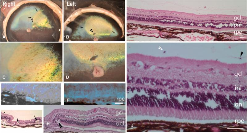
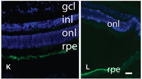
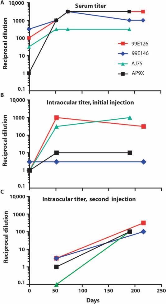
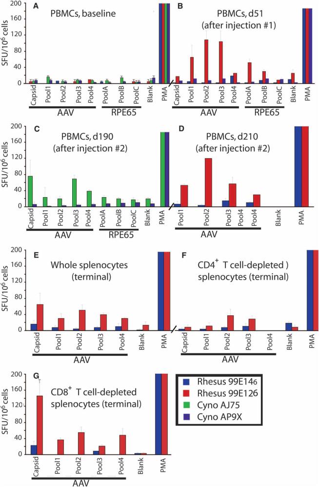
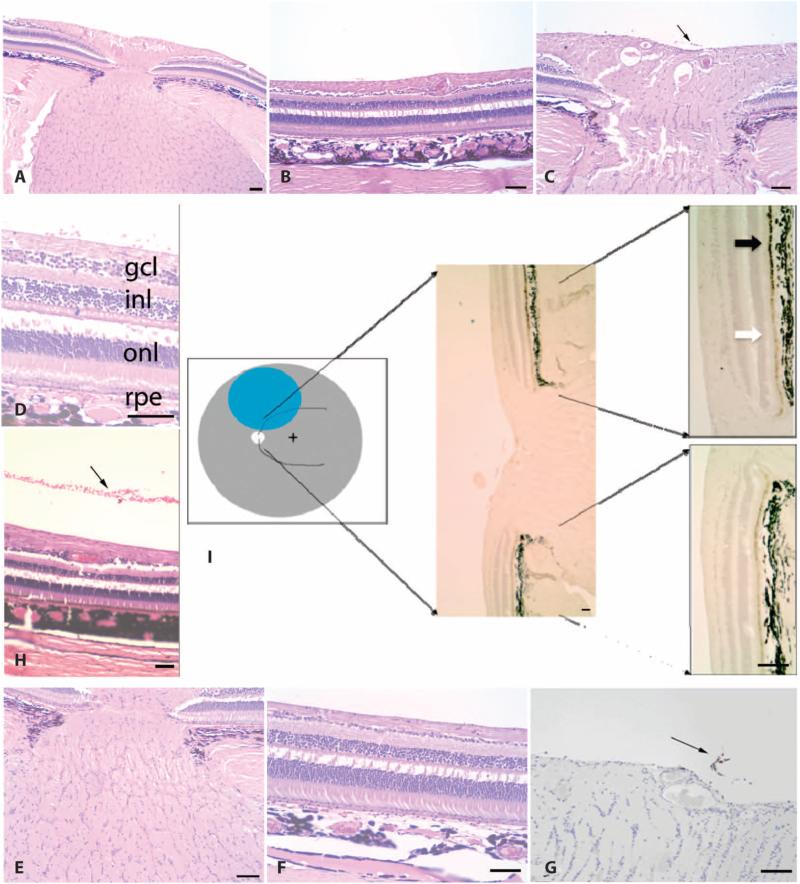
Leber's congenital amaurosis (LCA) is a group of severe inherited retinal degenerations that are symptomatic in infancy and lead to total blindness in adulthood. Recent clinical trials using recombinant adeno-associated virus serotype 2 (rAAV2) successfully reversed blindness in patients with LCA caused by RPE65 mutations after one subretinal injection. However, it was unclear whether treatment of the second eye in the same manner would be safe and efficacious, given the potential for a complicating immune response after the first injection. Here, we evaluated the immunological and functional consequences of readministration of rAAV2-hRPE65v2 to the contralateral eye using large animal models. Neither RPE65-mutant (affected; RPE65(-/-)) nor unaffected animals developed antibodies against the transgene product, but all developed neutralizing antibodies against the AAV2 capsid in sera and intraocular fluid after subretinal injection. Cell-mediated immune responses were benign, with only 1 of 10 animals in the study developing a persistent T cell immune response to AAV2, a response that was mediated by CD4(+) T cells. Sequential bilateral injection caused minimal inflammation and improved visual function in affected animals. Thus, subretinal readministration of rAAV2 in animals is safe and effective, even in the setting of preexisting immunity to the vector, a parameter that has been used to exclude patients from gene therapy trials.


Figures

Fig. 1
Fig. 1
Postmortem and histopathological analyses in affected dogs. (A to D) Borders of the original retinal detachment (arrows) and the retinotomy site (arrowhead) are apparent. (C and D) Higher-magnification views of (A) and (B), respectively. Shown are the right and left retinas of dog PE 810 days after injection. (E and F) Vacuoles are present in RPE cells from untreated (E) but not treated (F) portions of the retina (shown is dog SE, left eye). (G and H) Rosettes (arrows) and scarring (arrowhead) near the retinotomy sites (dog NE, left eye). (I and J) Normal retinal anatomy or layers. Plasma cells [arrowhead, identity confirmed in high-magnification images of (I) such as that shown in (J)] in the vitreous from dog NY (right eye). (K and L) RPE65 protein immunofluorescence (green) in AAV-treated (K, left half of L) but not untreated (right half of L) RPE. (K) Dog NY (right eye). (L) Dog NY (left eye). Scale bars, 100 mm. Nuclei are stained blue. gcl, ganglion cell layer; inl, inner nuclear layer; onl, outer nuclear layer.
Fig. 1
Fig. 1
Postmortem and histopathological analyses in affected dogs. (A to D) Borders of the original retinal detachment (arrows) and the retinotomy site (arrowhead) are apparent. (C and D) Higher-magnification views of (A) and (B), respectively. Shown are the right and left retinas of dog PE 810 days after injection. (E and F) Vacuoles are present in RPE cells from untreated (E) but not treated (F) portions of the retina (shown is dog SE, left eye). (G and H) Rosettes (arrows) and scarring (arrowhead) near the retinotomy sites (dog NE, left eye). (I and J) Normal retinal anatomy or layers. Plasma cells [arrowhead, identity confirmed in high-magnification images of (I) such as that shown in (J)] in the vitreous from dog NY (right eye). (K and L) RPE65 protein immunofluorescence (green) in AAV-treated (K, left half of L) but not untreated (right half of L) RPE. (K) Dog NY (right eye). (L) Dog NY (left eye). Scale bars, 100 mm. Nuclei are stained blue. gcl, ganglion cell layer; inl, inner nuclear layer; onl, outer nuclear layer.
Fig. 2
Fig. 2
NAbs in four NHPs directed against the AAV2 capsid. (A to C) NAbs in serum (A), AC fluid samples from the first (right) eye that was injected (B), and AC fluid samples from the second (left) eye that was injected (C). Days, days after first injection.
Fig. 3
Fig. 3
T cell responses directed against the AAV2 capsid or the RPE65 protein after subretinal readministration of AAV2-hRPE65v2 in NHPs. (A to D) IFN-γ secretion by PBMCs after stimulation with the AAV2 capsid and the RPE65 protein as measured by an IFN-γ ELISpot assay using peptide pools (see Supplementary Material). (A) Baseline. (B) Day 51 (d51). (C) Day 190 (d190). (D) Day 210 (d210). (E to G) IFN-γ ELISpot assay in whole splenocytes collected at necropsy (E), and CD4+ or CD8+ T cell–depleted splenocytes (F and G, respectively) in terminal samples from NHP 99E126. PMA, positive control. The error bars indicate SD.
Fig. 4
Fig. 4
(A to I) Histopathological (A to H) and immunohistochemical (I) analyses in retinas of unaffected NHPs after subretinal injection. Retinal layers (A to D, F, H, and I) and optic nerves (A, C, E, G, and I) have normal thicknesses. Mild injection-related changes include inflammatory cells (C, D, and G) and, in some, red blood cells in the vitreous (D and H). (A to F and H) Stained with H&E. (G) Stained with leukocyte common antigen. (I) Cartoon corresponding to the location of the subretinal injection (blue) with representative RPE65 immunohistochemical results shown at two magnifications. Black arrow, high levels of RPE65 protein in injected portion of the retina; white arrow, endogenous levels of RPE65 protein in unexposed retina. Scale bars, 100 mm. (A, B, E, and F) NHP 99E126 (right eye). (H) NHP 99E146 (right eye). (C, D, G, and I) NHP 99E126 (left eye). (B, F, D, and H) Processing artifacts cause a break between the inner and the outer nuclear layers (inl and onl, respectively). rpe, retinal pigment epithelium.

References

    1. Aleman TS, Jacobson SG, Chico JD, Scott ML, Cheung AY, Windsor EA, Furushima M, Redmond TM, Bennett J, Palczewski K, Cideciyan AV. Impairment of the transient pupillary light reflex in Rpe65−/− mice and humans with Leber congenital amaurosis. Invest. Ophthalmol. Vis. Sci. 2004;45:1259–1271. - PubMed
    1. Lorenz B, Gyürüs P, Preising M, Bremser D, Gu S, Andrassi M, Gerth C, Gal A. Early-onset severe rod-cone dystrophy in young children with RPE65 mutations. Invest. Ophthalmol. Vis. Sci. 2000;41:2735–2742. - PubMed
    1. Simonelli F, Ziviello C, Testa F, Rossi S, Fazzi E, Bianchi PE, Fossarello M, Signorini S, Bertone C, Galantuomo S, Brancati F, Valente EM, Ciccodicola A, Rinaldi E, Auricchio A, Banfi S. Clinical and molecular genetics of Leber’s congenital amaurosis: A multicenter study of Italian patients. Invest. Ophthalmol. Vis. Sci. 2007;48:4284–4290. - PubMed
    1. Perrault I, Rozet JM, Gerber S, Ghazi I, Leowski C, Ducroq D, Souied E, Dufier JL, Munnich A, Kaplan J. Leber congenital amaurosis. Mol. Genet. Metab. 1999;68:200–208. - PubMed
    1. den Hollander AI, Roepman R, Koenekoop RK, Cremers FP. Leber congenital amaurosis: Genes, proteins and disease mechanisms. Prog. Retin. Eye Res. 2008;27:391–419. - PubMed

Publication types

MeSH terms