Comparative Study
. 2010 Apr;133(Pt 4):973-82.
doi: 10.1093/brain/awq044.

A new alternative mechanism in glioblastoma vascularization: tubular vasculogenic mimicry

Affiliations
Comparative Study

A new alternative mechanism in glioblastoma vascularization: tubular vasculogenic mimicry

Soufiane El Hallani et al. Brain. 2010 Apr.

Abstract

Glioblastoma is one of the most angiogenic human tumours and endothelial proliferation is a hallmark of the disease. A better understanding of glioblastoma vasculature is needed to optimize anti-angiogenic therapy that has shown a high but transient efficacy. We analysed human glioblastoma tissues and found non-endothelial cell-lined blood vessels that were formed by tumour cells (vasculogenic mimicry of the tubular type). We hypothesized that CD133+ glioblastoma cells presenting stem-cell properties may express pro-vascular molecules allowing them to form blood vessels de novo. We demonstrated in vitro that glioblastoma stem-like cells were capable of vasculogenesis and endothelium-associated genes expression. Moreover, a fraction of these glioblastoma stem-like cells could transdifferentiate into vascular smooth muscle-like cells. We describe here a new mechanism of alternative glioblastoma vascularization and open a new perspective for the antivascular treatment strategy.


Figures
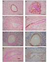
Figure 1
Figure 1
Non-endothelial blood vessels in glioblastoma. Endothelial cells are detected with anti-CD34 immunohistochemistry staining (dark brown) and vascular basement membrane with PAS staining (purple magenta) in (A) tubular blood vessel and (B) microvascular proliferation of glioblastomas. (C) Large vessel containing red blood cells stained positively with PAS but negatively with CD34. (D) Longitudinally sectioned blood vessel presents distinctive CD34+ and CD34 portions. (E) and (G) CD34 cell-lined vessels stain with collagen IV in the respective serial sections (F) and (H). Scale bars are 50 µm.
Figure 2
Figure 2
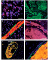
The tubular type vasculogenic mimicry in glioblastoma. (A) Nuclei are stained with DAPI (blue), fluorescent in situ hybridization EGFR probe (red) label tumour cells carrying EGFR amplification as double minutes (display multiple red signals). (B) Vascular smooth muscle cells are detected by anti-α-SMA immunofluorescence staining. (C) Nuclei of the vessel luminal surface do not harbour EGFR amplification. (D) Tumour cells carrying EGFR amplifications are visible in the luminal surface of a glioblastoma vessel. (E) The wall of an EGFR amplified cell-lined blood vessel presents α-SMA staining. Details of area delineated in (E) are enlarged in (F). Scale bars are 25 µm.
Figure 3
Figure 3
Characterization of glioblastoma stem-like cells. (A) Phase contrast (a) and fluorescent (b) microscopy of undifferentiated green fluorescent protein-expresing GSC growing into tumour spheres in neurosphere medium. Phase contrast microscopy of differentiated GSC at the differentiation assay (c). Scale bars are 100 µm. (B) Comparative genomic hybridization of GSC demonstrating tumour genomic alterations. Each bacterial artificial chromosome spotted on the comparative genomic hybridization array is represented by a dot. bacterial artificial chromosomes are ordered on the x-axis according to their position in the genome. For each chromosome the telomere of the short arm is on the left and the telomere of the long arm is on the right. The y-axis corresponds to fluorescence ratio. Yellow, green and red indicate genomic copy number normal, loss and gain, respectively. Genetic alteration includes complete chromosome 10 loss, gain of chromosome 7 with EGFR amplification (arrow). (C) Immunostaining of tumour cells for neural stem-cell markers (Nestin and Sox2) in the sphere, then astrocytic [glial fibrillary acidic protein (GFAP) in green staining] and neuronal (neuronal class III beta-tubulin-1 in red staining) markers by the differentiated cells around tumour sphere at Day 7. Scale bars are 50 µm. (D) Expression of Nestin, glial fibrillary acidic protein, neuronal class III beta-tubulin-1 and Tubulin by undifferentiated (sphere) and differentiated (diff) GSC were analysed by western blot as described. Extracts from primary rat astrocytes (Astro) and U87 were used as controls. TUJ-1 = neuronal class III beta-tubulin-1.
Figure 4
Figure 4
Glioblastoma stem-like cell vasculogenesis. (A) GSC-A and GSC-B are labelled with green fluorescent protein. GSC-A forms tubular structures in 3D culture on Matrigel while green fluorescent protein-expressing GSC-B does not. Scale bars are 250 µm. (B) Green fluorescent protein labelled-GSC-A exhibits a flattened morphology and α-SMA immunofluorescence after differentiation. Scale bars are 20 µm. (C) Expression of endothelial and vascular smooth muscle cell-related genes is measured by semiquantitative reverse transcriptase polymerase chain reaction in GSC-A and GSC-B compared to human cerebral microvascular endothelial cells and human vascular smooth muscle cells. The housekeeping gene ALAS is used as control.

Comment in

References

    1. Ausprunk DH, Folkman J. Migration and proliferation of endothelial cells in preformed and newly formed blood vessels during tumor angiogenesis. Microvasc Res. 1977;14:53–65. - PubMed
    1. Bonnet D, Dick JE. Human acute myeloid leukemia is organized as a hierarchy that originates from a primitive hematopoietic cell. Nat Med. 1997;3:730–7. - PubMed
    1. Brat DJ, Van Meir EG. Glomeruloid microvascular proliferation orchestrated by VPF/VEGF: a new world of angiogenesis research. Am J Pathol. 2001;158:789–6. - PMC - PubMed
    1. Chang YS, di Tomaso E, McDonald DM, Jones R, Jain RK, Munn LL. Mosaic blood vessels in tumors: frequency of cancer cells in contact with flowing blood. Proc Natl Acad Sci USA. 2000;97:14608–13. - PMC - PubMed
    1. Culley B, Murphy J, Babaie J, Nguyen D, Pagel A, Rousselle P, et al. Laminin-5 promotes neurite outgrowth from central and peripheral chick embryonic neurons. Neurosci Lett. 2001;301:83–6. - PubMed

Publication types

MeSH terms