. 2010 Jun 1;21(11):1897-907.
doi: 10.1091/mbc.e10-02-0145. Epub 2010 Apr 7.

Essential role for the CRAC activation domain in store-dependent oligomerization of STIM1

Affiliations

Essential role for the CRAC activation domain in store-dependent oligomerization of STIM1

Elizabeth D Covington et al. Mol Biol Cell. .

Abstract

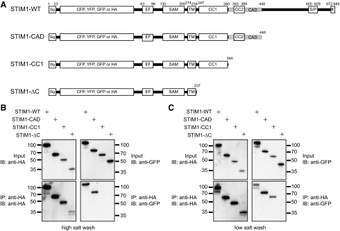
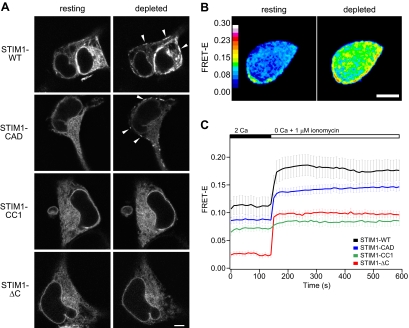
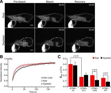
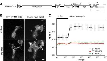
Oligomerization of the ER Ca(2+) sensor STIM1 is an essential step in store-operated Ca(2+) entry. The lumenal EF-hand and SAM domains of STIM1 are believed to initiate oligomerization after Ca(2+) store depletion, but the contributions of STIM1 cytosolic domains (coiled-coil 1, CC1; coiled-coil 2, CC2; CRAC activation domain, CAD) to this process are not well understood. By applying coimmunoprecipitation and fluorescence photobleaching and energy transfer techniques to truncated and mutant STIM1 proteins, we find that STIM1 cytosolic domains play distinct roles in forming both "resting" oligomers in cells with replete Ca(2+) stores and higher-order oligomers in store-depleted cells. CC1 supports the formation of resting STIM1 oligomers and appears to interact with cytosolic components to slow STIM1 diffusion. On store depletion, STIM1 lacking all cytosolic domains (STIM1-DeltaC) oligomerizes through EF-SAM interactions alone, but these oligomers are unstable. Addition of CC1 + CAD, but not CC1 alone, enables the formation of stable store-dependent oligomers. Within the CAD, both CC2 and C-terminal residues contribute to oligomer formation. Our results reveal a new function for the CAD: in addition to binding and activating Orai1, it is directly involved in STIM1 oligomerization, the initial event triggering store-operated Ca(2+) entry.


Figures

Figure 1.
Figure 1.
The CC1 domain supports resting self-association of STIM1. (A) Schematic of wild-type STIM1 (STIM1-WT) and truncated STIM1 proteins used in this study, with the major known domains indicated. Premature stop codons were introduced after the indicated residues. Abbreviations: Sig, signal peptide; EF, EF-hand; SAM, sterile alpha motif; TM, transmembrane domain; CC1, coiled-coil 1; CC2, coiled-coil 2; CAD, CRAC activation domain; S/P, serine-proline–rich region; K, polybasic region. (B) Only STIM1 proteins containing CC1 + CAD remain self-associated under high salt conditions. Cells coexpressing GFP- and HA-labeled STIM1 constructs were lysed in buffer containing 150 mM NaCl, and immunoprecipitates were washed in buffer containing 500 mM NaCl. Whole cell lysates (top) and anti-HA immunoprecipitates (bottom) were fractionated by SDS-PAGE and blotted with anti-HA antibodies (left) or anti-GFP antibodies (right). Representative of four experiments. (C) STIM1-CC1 remains self-associated under low-salt conditions, but STIM1 lacking the entire C-terminus (ΔC) does not. Cells were lysed and immunoprecipitates washed in buffer containing 10 mM NaCl. Representative of five experiments. The amino acid numbering does not include the CFP, YFP, GFP, or HA tag sequences.
Figure 2.
Figure 2.
Store-dependent, higher-order oligomerization of STIM1 requires the CRAC activation domain (CAD). (A) Confocal images focused at the equator of HEK 293 cells expressing GFP-labeled STIM1 mutants. Resting cells were bathed in 2 mM Ca2+ Ringer's solution (left), and the same cells were store-depleted with 4 μM ionomycin in Ca2+-free Ringer's solution for 5 min (right). Arrowheads mark locations of puncta. Bar, 5 μm. (B) FRET-E images of a HEK 293 cell expressing CFP-STIM1 and YFP-STIM1 before (left) and after (right) store depletion with 1 μM ionomycin in Ca2+-free Ringer's solution for 7 min. Bar, 5 μm. (C) Time course of mean FRET-E (±SEM) from cells coexpressing CFP- and YFP-labeled STIM1-WT (black, n = 8), STIM1-CAD (blue, n = 9), STIM1-CC1 (green, n = 7), or STIM1-ΔC (red, n = 14). Extracellular solution changes are indicated above the graph.
Figure 3.
Figure 3.
Diffusional mobility of truncated STIM1 mutants before and after store depletion. (A) FRAP in a HEK 293 cell expressing GFP-STIM1-CAD. After acquisition of prebleach images (left) a 3-μm-wide strip (indicated by the white rectangle, left) was photobleached (middle), and recovery was monitored over several minutes (right). FRAP was performed on the same cell again after store depletion with 0 mM Ca2+ Ringer + 4 μM ionomycin (bottom). Bar, 5 μm; time stamp, min:sec. (B) FRAP time course from the cell in A at rest (red) and after store depletion (black). Fits indicate a Deff of 0.23 μm2/s (rest) and 0.08 μm2/s (depleted). (C) Mean effective diffusion coefficients (±SEM) before (red) or after (black) treatment with ionomycin to deplete Ca2+ stores. Numbers of cells (rest, depleted): STIM1-ΔC (n = 8, 6); STIM1-CC1 (n = 10, 9); STIM1-CAD (n = 8, 13); STIM1-WT (n = 32, 9). Significant differences, determined by Student's t tests, between mean Deff are noted by p values.
Figure 4.
Figure 4.
Mutations in the CC2 domain of CAD identify residues critical for STIM1 function and oligomerization. (A–D) Confocal images of the footprints of HEK 293 cells cotransfected with mCherry-myc-Orai1 and GFP-labeled STIM1-WT (A), STIM1-L373K (B), STIM1-A369K (C), or STIM1-A376K (D). In each panel the same cell is displayed before and after treatment with 1 μM TG to deplete stores. Bars, 5 μm. (E) Effects of STIM1 CC2 mutations on SOCE activation. Mean Fura-2 ratios (±SEM) are shown for HEK 293 cells cotransfected with mCherry-myc-Orai1 and STIM1-WT (black, n = 32), STIM1-L373K (blue, n = 29), STIM1-A376K (red, n = 21), or STIM1-A369K (green, n = 33). Stores were depleted with 1 μM TG, and extracellular Ca2+ concentration in mM is indicated above the graph. (F) Time course of mean whole cell FRET-E (±SEM) from HEK 293 cells expressing CFP- and YFP-labeled STIM1-WT (black, n = 8), STIM1-L373K (blue, n = 16), STIM1-A376K (red, n = 9), or STIM1-A369K (green; analysis of cell periphery only, green circles; n = 9). Stores were depleted with 1 μM ionomycin in Ca2+-free Ringer's solution as indicated.
Figure 5.
Figure 5.
STIM1-CC2 fails to oligomerize in response to store depletion. (A) STIM1-CC2 was constructed by introducing a premature stop codon at aa 392 as shown. (B) Confocal images of the footprint of a HEK 293 cell cotransfected with CFP-STIM1-CC2 and mCherry-myc-Orai1, before and after treatment with 1 μM TG to deplete Ca2+ stores. Bar, 5 μm. (C) Time course of mean FRET-E (±SEM) from HEK 293 cells coexpressing CFP- and YFP-labeled STIM1-CC2 (red, n = 9); the FRET-E response of STIM1-WT (black, n = 8) and STIM1-CC1 (green, n = 7) are reproduced from Figure 2C for comparison. 1 μM ionomycin was added as indicated above the graph.

Similar articles

Cited by

References

    1. Baba Y., et al. Coupling of STIM1 to store-operated Ca2+ entry through its constitutive and inducible movement in the endoplasmic reticulum. Proc. Natl. Acad. Sci .USA. 2006;103:16704–16709. - PMC - PubMed
    1. Bautista D. M., Hoth M., Lewis R. S. Enhancement of calcium signalling dynamics and stability by delayed modulation of the plasma-membrane calcium-ATPase in human T cells. J. Physiol. 2002;541:877–894. - PMC - PubMed
    1. Berney C., Danuser G. FRET or no FRET: a quantitative comparison. Biophys. J. 2003;84:3992–4010. - PMC - PubMed
    1. Blanchetot C., Tertoolen L. G., den Hertog J. Regulation of receptor protein-tyrosine phosphatase alpha by oxidative stress. EMBO J. 2002;21:493–503. - PMC - PubMed
    1. Cahalan M. D. STIMulating store-operated Ca2+ entry. Nat. Cell Biol. 2009;11:669–677. - PMC - PubMed

Publication types

MeSH terms

LinkOut - more resources