. 2010 Jun 1;21(11):1885-96.
doi: 10.1091/mbc.e09-12-1041. Epub 2010 Apr 7.

The mobile FG nucleoporin Nup98 is a cofactor for Crm1-dependent protein export

Affiliations

The mobile FG nucleoporin Nup98 is a cofactor for Crm1-dependent protein export

Masahiro Oka et al. Mol Biol Cell. .

Abstract

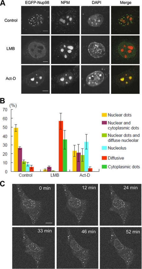
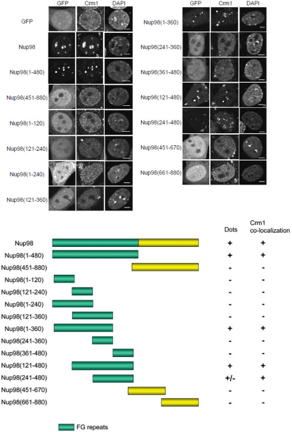
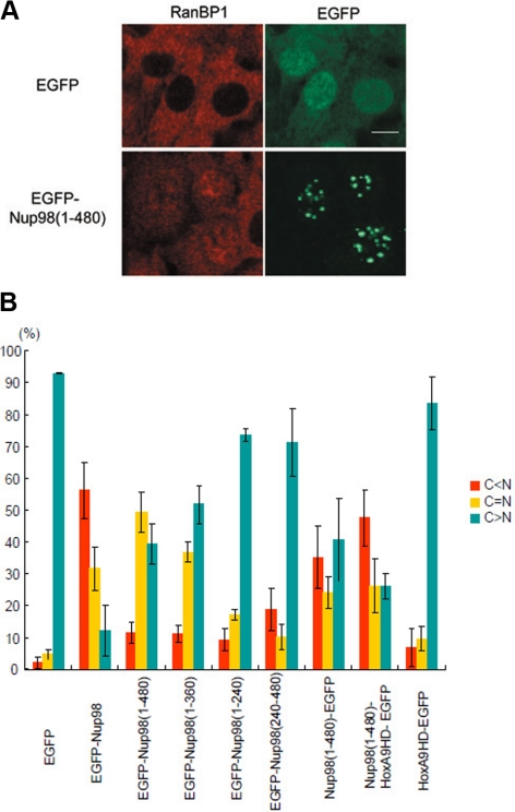
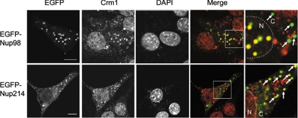
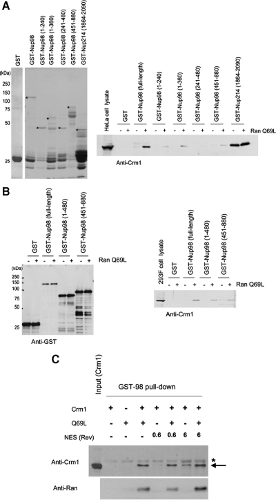
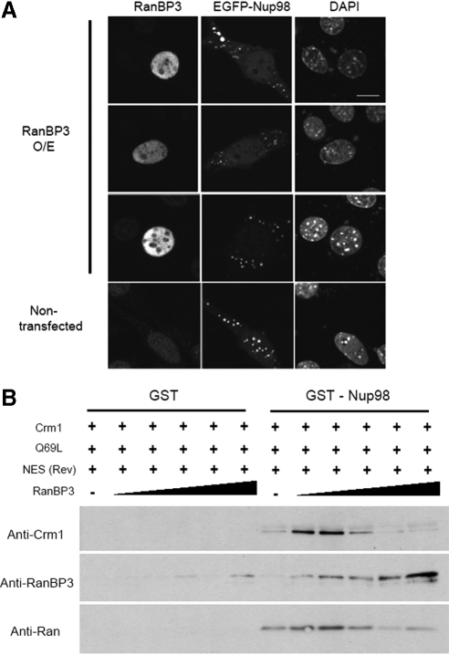
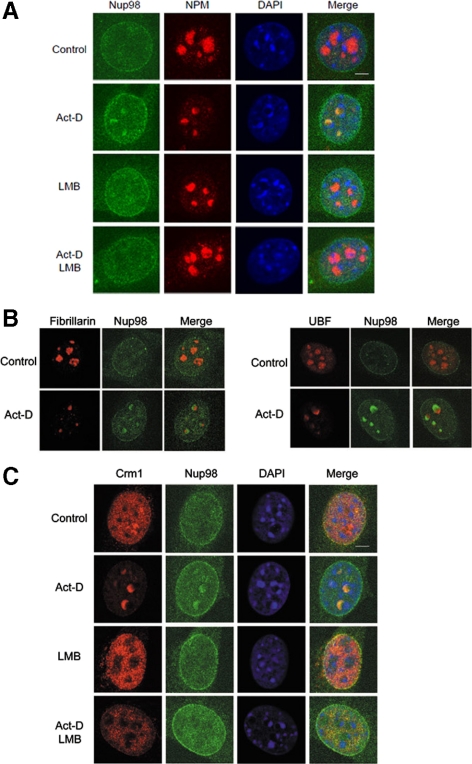
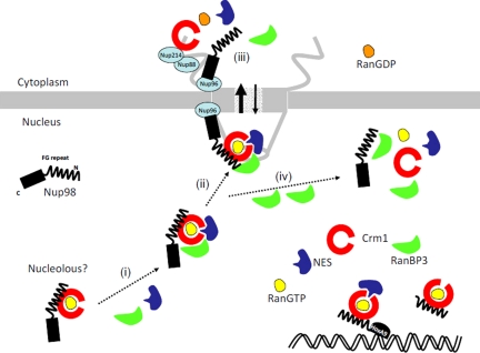
Nup98 is a mobile nucleoporin that forms distinct dots in the nucleus, and, although a role for Nup98 in nuclear transport has been suggested, its precise function remains unclear. Here, we show that Nup98 plays an important role in Crm1-mediated nuclear protein export. Nuclear, but not cytoplasmic, dots of EGFP-tagged Nup98 disappeared rapidly after cell treatment with leptomycin B, a specific inhibitor of the nuclear export receptor, Crm1. Mutational analysis demonstrated that Nup98 physically and functionally interacts with Crm1 in a RanGTP-dependent manner through its N-terminal phenylalanine-glycine (FG) repeat region. Moreover, the activity of the Nup98-Crm1 complex was modulated by RanBP3, a known cofactor for Crm1-mediated nuclear export. Finally, cytoplasmic microinjection of anti-Nup98 inhibited the Crm1-dependent nuclear export of proteins, concomitant with the accumulation of anti-Nup98 in the nucleus. These results clearly demonstrate that Nup98 functions as a novel shuttling cofactor for Crm1-mediated nuclear export in conjunction with RanBP3.


Figures

Figure 1.
Figure 1.
EGFP-Nup98 dots redistribute after LMB or Act-D treatment. (A) NIH 3T3 cells were transfected with EGFP-Nup98 expression vector. After 48 h, the cells were treated with DMSO (Control), 5 nM LMB, or 40 ng/ml Act-D for 2 h. Cells were then fixed, immunostained, and examined by confocal microscopy. Anti-nucleophosmin/B23 (NPM) antibody was used to stain the nucleolus. DAPI staining was used to visualize nuclei. Bar, 10 μm. (B) Summary of the results obtained in A. At least 150 EGFP-Nup98–expressing cells were observed for each condition. The localization patterns of EGFP-Nup98 were categorized into six groups as follows: nuclear dots, nuclear and cytoplasmic dots, cytoplasmic dots and diffuse nuclear staining, nuclear dots and diffuse nucleolar staining, diffuse nucleolar staining, and diffuse nuclear staining. Data are represented as mean ± SD of three independent experiments. (C) Time-lapse analysis of EGFP-Nup98 dots after LMB treatment. EGFP-Nup98–expressing NIH3T3 cells were treated with 5 nM LMB and analyzed by time-lapse microscopy. The position of the nucleus is indicated by a dashed circle. Bar, 10 μm.
Figure 2.
Figure 2.
The N-terminal FG-repeat region of Nup98 is required for the formation of nuclear dots that colocalize with Crm1. NIH 3T3 cells were transfected with EGFP-Nup98 or the indicated EGFP-Nup98 deletion mutants. After 48 h, the cells were fixed, permeabilized, and immunostained with anti-Crm1 antibody. The formation of nuclear dots and the localization of Crm1 were determined. DAPI staining was used to visualize nuclei. Bar, 5 μm.
Figure 3.
Figure 3.
The FG-repeat region of Nup98 affects the function of Crm1. (A) NIH 3T3 cells were transfected with EGFP or EGFP-Nup98 (aa 1-480) expression vectors. After 48 h, the cells were immunostained with anti-RanBP1 antibody. Bar, 10 μm. (B) NIH 3T3 cells were transfected with the indicated expression vectors. After 48 h, the cells were immunostained with anti-RanBP1, and the cellular localization of RanBP1 in EGFP-positive cells was categorized as C > N, C = N, or C < N. Data are represented as mean ± SD of three independent experiments.
Figure 4.
Figure 4.
Nuclear, but not cytoplasmic, Nup98 dots colocalize with Crm1. NIH 3T3 cells were transfected with EGFP-Nup98– or EGFP-Nup214– encoding plasmids. After 48 h, the cells were fixed, permeabilized, and immunostained with anti-Crm1. DAPI staining was used to visualize nuclei. Arrows indicate the EGFP-Nup dots in the cytoplasm. Merged images of EGFP-Nup98 or EGFP-Nup214 (green) and Crm1 (red) are shown. N, nucleus; C, cytoplasm. Bar, 10 μm.
Figure 5.
Figure 5.
Nup98 associates with Crm1 in a Ran-GTP–dependent manner. (A) Left, bacterially expressed GST, GST-Nup98 (full-length or deletion mutants), and GST-Nup214 FG-repeat region (1864-2090) were purified and analyzed by SDS-PAGE and Coomassie staining. Right, purified GST or GST-fusion proteins bound to glutathione beads were incubated with HeLa cell lysates (300 μg) in the presence or absence of 2 μM RanQ69L-GTP for 3 h at 4°C. The beads were washed, and the associated proteins were eluted and analyzed by immunoblotting using anti-Crm1. Thirty micrograms of whole cell lysate from HeLa cells was used for an input control. Asterisks indicate the full-length GST-fusion proteins. (B) GST-tagged Nup98 or deletion mutants were transiently expressed in HEK293F cells. The cell lysates (500 μg) were prepared and incubated with glutathione beads in the presence or absence of 2 μM RanQ69L-GTP for 3 h. The beads were washed, and associated proteins were eluted and analyzed by immunoblotting using anti-GST (left) or anti-Crm1 (right). Twenty-five micrograms of whole cell lysate from 293F cells was used for an input control. (C) Purified recombinant GST-Nup98 (5 μg) was incubated with the indicated mixtures of Crm1 (200 nM), RanQ69L (1 μM), NES peptide (Rev; 0.6–6 μM) for 2 h at 4°C, and precipitated using glutathione-Sepharose 4B beads. The bound proteins were analyzed by immunoblotting using anti-Crm1 or anti-Ran. Asterisk indicates GST-Nup98, which weakly cross-reacted with the anti-Crm1 antibody.
Figure 6.
Figure 6.
RanBP3 modulates the Nup98-Crm1 interaction in a biphasic manner. (A) Cells were transfected with EGFP-Nup98 alone or in combination with RanBP3 for 48 h. The cells were then fixed and examined by confocal microscopy. The nuclei were visualized by DAPI staining. Bar, 10 μm. (B) GST-Nup98 (5 μg) was incubated with Crm1 (200 nM), RanQ69L (1 μM), NES (6 μM), and different concentrations of RanBP3 (from left to right: 11, 33, 100, 300, and 900 nM) for 2 h at 4°C and was precipitated using glutathione-Sepharose 4B beads. The bound proteins were analyzed by immunoblotting.
Figure 7.
Figure 7.
Endogenous Nup98 relocalizes to the granular compartment of the nucleolus after Act-D treatment in a Crm1-dependent manner. (A) NIH 3T3 cells were treated with either Act-D or LMB, or both for 2 h. The cells were fixed, permeabilized, and immunostained with anti-nucleophosmin/B23 (NPM) and anti-Nup98. The nuclei were visualized by DAPI staining. Bar, 5 μm. (B) NIH 3T3 cells were treated with Act-D or DMSO (Control) for 2 h. The cells were fixed, permeabilized, and immunostained with anti-Nup98 together with the indicated antibody to stain the components of the nucleolus. Merged images are shown. Bar, 5 μm. (C) NIH 3T3 cells were treated with either Act-D or LMB, or both for 2 h. The cells were fixed, permeabilized, and immunostained with anti-Crm1 and anti-Nup98. The nuclei were visualized by DAPI staining. Bar, 5 μm.
Figure 8.
Figure 8.
Microinjection of anti-Nup98 inhibits NES-dependent nuclear export. (A) HeLa cells were microinjected with control IgG, anti-Nup62, or anti-Nup98 together with GST-GFP-NES and Alexa568-conjugated anti-sheep IgG (injection marker) into the nucleus. After 15 min of incubation at 37°C, the cells were fixed, washed, and mounted for microscopic observation. Bar, 20 μm. (B) HeLa cells were cytoplasmically microinjected with control IgG, anti-Nup62, or anti-Nup98. After 15 min of incubation at 37°C, the cells were fixed and stained with anti-RanBP1. Alexa488-conjugated anti-rat IgG or Alexa568-conjugated anti-rabbit IgG were used to detect rat monoclonal IgG or anti-RanBP1, respectively. Bar, 20 μm. (C) HeLa cells were microinjected with anti-Nup62 or anti-Nup98 in the cytoplasm, together with Alexa568-conjugated anti-sheep IgG (injection marker). After 15 min of incubation at 37°C, the cells were fixed and stained with Alexa488-conjugated anti-rat IgG. Bar, 20 μm.
Figure 9.
Figure 9.
A model for the role of Nup98 in Crm1-mediated nuclear protein export. Nup98 forms a complex with Crm1 in a RanGTP-dependent manner in the nucleus or nucleolus. RanBP3 facilitates the formation of the NES-cargo–containing Nup98-RanBP3-Crm1-RanGTP complex (i). This complex is then targeted to the NPC (ii) and transported into the cytoplasm. The C-terminal non-FG-repeat domain of Nup98 is involved in the targeting of the export complex to the NPC facilitating the Crm1-dependent translocation of NES-containing proteins through the NPC. After translocation, the export complex dissociates after the conversion of RanGTP to RanGDP (iii). When an excess amount of RanBP3 is present, the NES-cargo containing complex dissociates (iv). Both the FG-repeat fragment of Nup98 and Nup98-HoxA9 fusion interact with Crm1 but are not targeted to the NPC, and both inhibit the function of Crm1.

Similar articles

Cited by

References

    1. Bastos R., Ribas de Pouplana L., Enarson M., Bodoor K., Burke B. Nup84, a novel nucleoporin that is associated with CAN/Nup214 on the cytoplasmic face of the nuclear pore complex. J. Cell Biol. 1997;137:989–1000. - PMC - PubMed
    1. Belgareh N., et al. An evolutionarily conserved NPC subcomplex, which redistributes in part to kinetochores in mammalian cells. J. Cell Biol. 2001;154:1147–1160. - PMC - PubMed
    1. Ben-Efraim I., Gerace L. Gradient of increasing affinity of importin beta for nucleoporins along the pathway of nuclear import. J. Cell Biol. 2001;152:411–417. - PMC - PubMed
    1. Bischoff F. R., Klebe C., Kretschmer J., Wittinghofer A., Ponstingl H. RanGAP1 induces GTPase activity of nuclear Ras-related Ran. Proc. Natl. Acad. Sci. USA. 1994;91:2587–2591. - PMC - PubMed
    1. Boer J., Bonten-Surtel J., Grosveld G. Overexpression of the nucleoporin CAN/NUP214 induces growth arrest, nucleocytoplasmic transport defects, and apoptosis. Mol. Cell. Biol. 1998;18:1236–1247. - PMC - PubMed

Publication types

MeSH terms