Review
. 2010 Sep;84(17):8360-8.
doi: 10.1128/JVI.00443-10. Epub 2010 Apr 7.

Virus cell-to-cell transmission

Affiliations
Review

Virus cell-to-cell transmission

Walther Mothes et al. J Virol. 2010 Sep.

Abstract

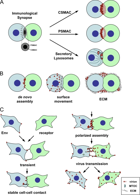
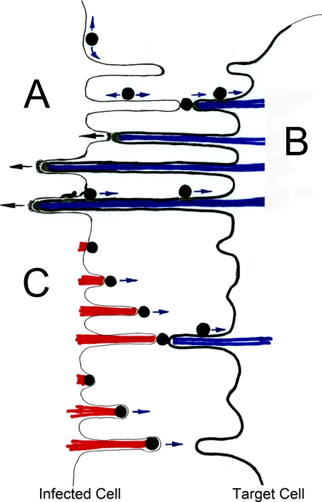
Viral infections spread based on the ability of viruses to overcome multiple barriers and move from cell to cell, tissue to tissue, and person to person and even across species. While there are fundamental differences between these types of transmissions, it has emerged that the ability of viruses to utilize and manipulate cell-cell contact contributes to the success of viral infections. Central to the excitement in the field of virus cell-to-cell transmission is the idea that cell-to-cell spread is more than the sum of the processes of virus release and entry. This implies that virus release and entry are efficiently coordinated to sites of cell-cell contact, resulting in a process that is distinct from its individual components. In this review, we will present support for this model, illustrate the ability of viruses to utilize and manipulate cell adhesion molecules, and discuss the mechanism and driving forces of directional spreading. An understanding of viral cell-to-cell spreading will enhance our ability to intervene in the efficient spreading of viral infections.


Figures

FIG. 1.
FIG. 1.
Viruses can either utilize existing synaptic contacts or establish contact between infected and uninfected cells to promote viral spreading. (A) Utilization of structural elements of immunological synapses for viral spreading. The architecture of the immunological synapse consists of central and peripheral supermolecular adhesion complexes, cSMAC and pSMAC, respectively (84). The antigen-presenting cell is shown in green, and the effector cell is shown in blue. Exocytosis of secretory lysosomes can be observed in the cSMAC zone. Interestingly, all three structural elements have been associated with the accumulation and transmission of viruses from infected to uninfected cells (43, 51, 102). (B) Several distinct mechanisms can contribute to the accumulation of viruses at the synapse. The association of viruses with the cell-cell interface could be the consequence of either de novo assembly or surface transmission (55, 56). Extracellular matrix components (ECM) have also been observed to play a role in a peripheral mode of transmission (92). Virus-infected cells are blue, and the receptor-expressing target cells are labeled as green. (C) Establishment of adhesion between infected and uninfected cells using MLV as the model system (56, 112). MLV-infected fibroblasts were observed to establish contact with uninfected cells in a reaction driven by Env-receptor interactions. Initial transient contacts are stabilized, and virus assembly was observed to be redirected toward sites of cell-cell contact after an ∼30-min delay. Following virus transmission, cell-cell contacts are downregulated. Virus-infected cells are blue, and the receptor-expressing target cells are green.
FIG. 2.
FIG. 2.
Driving forces for directional spreading of viruses (adapted from reference with permission of the publisher). (A) Affinity-based model for directional spreading. Downregulation of the viral receptor in the infected cells allows the establishment of a general affinity gradient between the infected and uninfected cell. Additional virus interactions with the infected cell can retain completely budded viral particles at the surface of the infected cell. Low-affinity retention on the surface of producer cells allows for diffusive movement along the cell surface (111). This process can position viruses for subsequent high-affinity interactions with target cells expressing receptor, thereby driving directional spreading of surface associated viruses. (B) Viral transmission driven by retrograde F-actin flow of the target cell. High-affinity interactions with receptor (green) in the target cell allows surface-associated viruses to engage the underlying F-actin flow (blue) to move toward the cell body of the target cell (17, 68, 69, 110, 115). This process, originally designated “virus surfing,” can promote a driving force for cell-to-cell spreading for the surface-retained viruses presented in panel A. In addition, long-term Env-receptor interactions between infected and uninfected cells can anchor target cell membranes directly at the infected cells (112). Because virus assembly can be redirected toward these sites of cell-cell contact (56), viruses can immediately engage target cell F-actin flow (blue) to move toward the target cell upon completion of assembly. (C) Viral transmission driven by actin assembly in the infected cell. In contrast to the depictions in panel B, viruses can induce actin comet tail formation (red) inside the infected cell to propel themselves toward neighboring cells (23, 30, 62). While vaccinia virus induces actin tails (red) beneath the plasma membrane of completely released viruses, capsids of the African swine fever virus induce actin tails (red) while still topologically within the cytoplasm. Such actin-propelled motion may be sufficient to drive transmission, but a combination of actin-driven movements both away from the infected cell (red) as well as toward the target cell (blue) are also plausible (81).

References

    1. Abe, K., O. Chisaka, F. Van Roy, and M. Takeichi. 2004. Stability of dendritic spines and synaptic contacts is controlled by alpha N-catenin. Nat. Neurosci. 7:357-363. - PubMed
    1. Agosto, L. M., J. J. Yu, M. K. Liszewski, C. Baytop, N. Korokhov, L. M. Humeau, and U. O'Doherty. 2009. The CXCR4-tropic human immunodeficiency virus envelope promotes more-efficient gene delivery to resting CD4+ T cells than the vesicular stomatitis virus glycoprotein G envelope. J. Virol. 83:8153-8162. - PMC - PubMed
    1. Akins, M. R., and T. Biederer. 2006. Cell-cell interactions in synaptogenesis. Curr. Opin. Neurobiol. 16:83-89. - PubMed
    1. Alfsen, A., H. Yu, A. Magerus-Chatinet, A. Schmitt, and M. Bomsel. 2005. HIV-1-infected blood mononuclear cells form an integrin- and agrin-dependent viral synapse to induce efficient HIV-1 transcytosis across epithelial cell monolayer. Mol. Biol. Cell 16:4267-4279. - PMC - PubMed
    1. Arhel, N., M. Lehmann, K. Clauss, G. U. Nienhaus, V. Piguet, and F. Kirchhoff. 2009. The inability to disrupt the immunological synapse between infected human T cells and APCs distinguishes HIV-1 from most other primate lentiviruses. J. Clin. Invest. 119:2965-2975. - PMC - PubMed

Publication types