

Novel recombinant Mycobacterium bovis BCG, ovine atadenovirus, and modified vaccinia virus Ankara vaccines combine to induce robust human immunodeficiency virus-specific CD4 and CD8 T-cell responses in rhesus macaques
- PMID: 20375158
- PMCID: PMC2876636
- DOI: 10.1128/JVI.02607-09
Novel recombinant Mycobacterium bovis BCG, ovine atadenovirus, and modified vaccinia virus Ankara vaccines combine to induce robust human immunodeficiency virus-specific CD4 and CD8 T-cell responses in rhesus macaques
Abstract
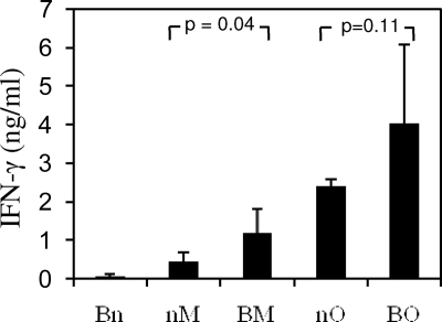
Mycobacterium bovis bacillus Calmette-Guérin (BCG), which elicits a degree of protective immunity against tuberculosis, is the most widely used vaccine in the world. Due to its persistence and immunogenicity, BCG has been proposed as a vector for vaccines against other infections, including HIV-1. BCG has a very good safety record, although it can cause disseminated disease in immunocompromised individuals. Here, we constructed a recombinant BCG vector expressing HIV-1 clade A-derived immunogen HIVA using the recently described safer and more immunogenic BCG strain AERAS-401 as the parental mycobacterium. Using routine ex vivo T-cell assays, BCG.HIVA(401) as a stand-alone vaccine induced undetectable and weak CD8 T-cell responses in BALB/c mice and rhesus macaques, respectively. However, when BCG.HIVA(401) was used as a priming component in heterologous vaccination regimens together with recombinant modified vaccinia virus Ankara-vectored MVA.HIVA and ovine atadenovirus-vectored OAdV.HIVA vaccines, robust HIV-1-specific T-cell responses were elicited. These high-frequency T-cell responses were broadly directed and capable of proliferation in response to recall antigen. Furthermore, multiple antigen-specific T-cell clonotypes were efficiently recruited into the memory pool. These desirable features are thought to be associated with good control of HIV-1 infection. In addition, strong and persistent T-cell responses specific for the BCG-derived purified protein derivative (PPD) antigen were induced. This work is the first demonstration of immunogenicity for two novel vaccine vectors and the corresponding candidate HIV-1 vaccines BCG.HIVA(401) and OAdV.HIVA in nonhuman primates. These results strongly support their further exploration.
Figures


Similar articles
-
Safety and immunogenicity of novel recombinant BCG and modified vaccinia virus Ankara vaccines in neonate rhesus macaques.J Virol. 2010 Aug;84(15):7815-21. doi: 10.1128/JVI.00726-10. Epub 2010 May 19. J Virol. 2010. PMID: 20484495 Free PMC article.
-
Optimizing HIV-1-specific CD8+ T-cell induction by recombinant BCG in prime-boost regimens with heterologous viral vectors.Eur J Immunol. 2011 Dec;41(12):3542-52. doi: 10.1002/eji.201141962. Epub 2011 Oct 26. Eur J Immunol. 2011. PMID: 21932450
-
Preclinical development of BCG.HIVA2auxo.int, harboring an integrative expression vector, for a HIV-TB Pediatric vaccine. Enhancement of stability and specific HIV-1 T-cell immunity.Hum Vaccin Immunother. 2017 Aug 3;13(8):1798-1810. doi: 10.1080/21645515.2017.1316911. Epub 2017 Apr 20. Hum Vaccin Immunother. 2017. PMID: 28426273 Free PMC article.
-
Development of a DNA-MVA/HIVA vaccine for Kenya.Vaccine. 2002 May 6;20(15):1995-8. doi: 10.1016/s0264-410x(02)00085-3. Vaccine. 2002. PMID: 11983261 Review.
-
Clinical experience with plasmid DNA- and modified vaccinia virus Ankara-vectored human immunodeficiency virus type 1 clade A vaccine focusing on T-cell induction.J Gen Virol. 2007 Jan;88(Pt 1):1-12. doi: 10.1099/vir.0.82493-0. J Gen Virol. 2007. PMID: 17170430 Review.
Cited by
-
Safety and immunogenicity of novel recombinant BCG and modified vaccinia virus Ankara vaccines in neonate rhesus macaques.J Virol. 2010 Aug;84(15):7815-21. doi: 10.1128/JVI.00726-10. Epub 2010 May 19. J Virol. 2010. PMID: 20484495 Free PMC article.
-
Priming with Recombinant BCG Expressing HTI Enhances the Magnitude and Breadth of the T-Cell Immune Responses Elicited by MVA.HTI in BALB/c Mice.Vaccines (Basel). 2020 Nov 13;8(4):678. doi: 10.3390/vaccines8040678. Vaccines (Basel). 2020. PMID: 33202884 Free PMC article.
-
Priming with recombinant auxotrophic BCG expressing HIV-1 Gag, RT and Gp120 and boosting with recombinant MVA induces a robust T cell response in mice.PLoS One. 2013 Aug 20;8(8):e71601. doi: 10.1371/journal.pone.0071601. eCollection 2013. PLoS One. 2013. PMID: 23977084 Free PMC article.
-
Dual neonate vaccine platform against HIV-1 and M. tuberculosis.PLoS One. 2011;6(5):e20067. doi: 10.1371/journal.pone.0020067. Epub 2011 May 13. PLoS One. 2011. PMID: 21603645 Free PMC article.
-
Exploring HIV Vaccine Progress in the Pre-Clinical and Clinical Setting: From History to Future Prospects.Viruses. 2024 Feb 27;16(3):368. doi: 10.3390/v16030368. Viruses. 2024. PMID: 38543734 Free PMC article. Review.
References
-
- Barouch, D. H., M. G. Pau, J. H. Custers, W. Koudstaal, S. Kostense, M. J. Havenga, D. M. Truitt, S. M. Sumida, M. G. Kishko, J. C. Arthur, B. Korioth-Schmitz, M. H. Newberg, D. A. Gorgone, M. A. Lifton, D. L. Panicali, G. J. Nabel, N. L. Letvin, and J. Goudsmit. 2004. Immunogenicity of recombinant adenovirus serotype 35 vaccine in the presence of pre-existing anti-Ad5 immunity. J. Immunol. 172:6290-6297. - PubMed
-
- Both, G. W. 2004. Ovine atadenovirus: a review of its biology, biosafety profile and application as a gene delivery vector. Immunol. Cell Biol. 82:189-195. - PubMed
-
- Both, G. W., F. Cameron, A. Collins, L. J. Lockett, and J. Shaw. 2007. Production and release testing of ovine atadenovirus vectors. Methods Mol. Med. 130:69-90. - PubMed
-
- Brandt, L., Y. A. Skeiky, M. R. Alderson, Y. Lobet, W. Dalemans, O. C. Turner, R. J. Basaraba, A. A. Izzo, T. M. Lasco, P. L. Chapman, S. G. Reed, and I. M. Orme. 2004. The protective effect of the Mycobacterium bovis BCG vaccine is increased by coadministration with the Mycobacterium tuberculosis 72-kilodalton fusion polyprotein Mtb72F in M. tuberculosis-infected guinea pigs. Infect. Immun. 72:6622-6632. - PMC - PubMed
-
- Bridgeman, A., Y. Roshorm, L. J. Lockett, Z. Z. Xu, R. Hopkins, J. Shaw, G. W. Both, and T. Hanke. 2010. Ovine atadenovirus, a novel and highly immunogenic vector in prime-boost studies of a candidate HIV-1 vaccine. Vaccine 28:474-483. - PubMed
Publication types
MeSH terms
Substances
Grants and funding
LinkOut - more resources
Full Text Sources
Medical
Research Materials
