

doi: 10.1128/JVI.02375-09.
Epub 2010 Apr 7.
Cell entry of the aphthovirus equine rhinitis A virus is dependent on endosome acidification
Affiliations
- PMID: 20375159
- PMCID: PMC2876639
- DOI: 10.1128/JVI.02375-09
Item in Clipboard
Cell entry of the aphthovirus equine rhinitis A virus is dependent on endosome acidification
J Virol.
2010 Jun.
Abstract
Equine rhinitis A virus (ERAV) is genetically closely related to foot-and-mouth disease virus (FMDV), and both are now classified within the genus Aphthovirus of the family Picornaviridae. For disease security reasons, FMDV can be handled only in high-containment facilities, but these constraints do not apply to ERAV, making it an attractive alternative for the study of aphthovirus biology. Here, we show, using immunofluorescence, pharmacological agents, and dominant negative inhibitors, that ERAV entry occurs (as for FMDV) via clathrin-mediated endocytosis and acidification of early endosomes. This validates the use of ERAV as a model system to study the mechanism of cell entry by FMDV.
Figures
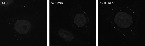
ERAV internalization time course. ERAV-Cy2 was bound to cells in the cold, and unbound virus was washed away before infection initiated at physiological temperature. Cells were fixed at 0, 5, and 10 min pi (panels a, b, and c, respectively).
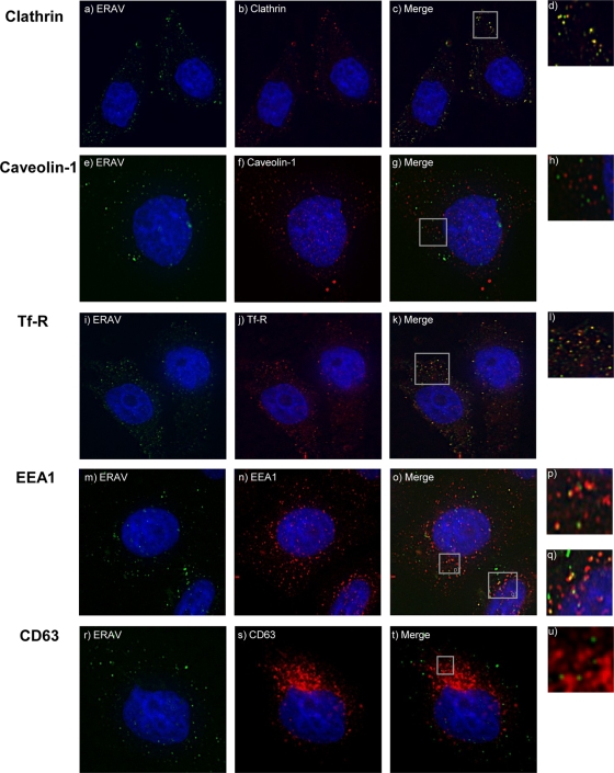
ERAV colocalization with endocytosis markers. Cells were infected with Cy2-labeled ERAV for 0 to 30 min before being visualized as described in the text. Antibodies against endocytosis markers were used in combination with secondary antibodies conjugated to Alexa-594 (Invitrogen). Cell nuclei were stained with Hoechst. Images were acquired using a DeltaVision deconvoluting microscope. (a) ERAV-Cy2 (green). (b) Clathrin (red) at 10 min pi. (c) Merge of panels a and b, showing colocalization (yellow) of ERAV and clathrin. (e to t) Cells fixed at 20 min pi with ERAV-Cy2 (green) and caveolin-1, Tfr-R, EEA1, or CD63 (red). (g) Overlay of panels e and f, showing ERAV (green) and caveolin-1 (red). (k and o) Colocalization (yellow) of ERAV with Tfr-R or EEA1, respectively. Panel k is a merge of panels i and j, and panel o is a merge of panels m and n. (t) Overlay of panels r and s, showing ERAV (green) and CD63 (red). Panels d, h, l, p, q, and u are enlargements from the Merge panels.

Effects of inhibition of clathrin-mediated endocytosis on ERAV entry. (A) Detection of ERAV (red) 20 min pi (b and f) of cells expressing wild-type Eps15 (Eps15-WT-GFP) (green) (a) or dominant negative Eps15 (Eps15-DN-GFP) (green) (e), which inhibits clathrin-mediated endocytosis. ERAV is located predominantly in the cytoplasm in cells expressing wild-type Eps15 (c, overlay of a and b). Panel d is an enlargement from panel c. ERAV is located predominantly at the cell surface in cells expressing dominant negative Eps15 (g, overlay of panels e and f). Panel h is an enlargement from panel g. (B) Panels i to o show cells fixed at 6 h pi. Panel k is a merge of panels i (Eps15-WT-GFP [green]) and j (ERAV [red]). The large ERAV signal in the cytoplasm is indicative of replication. Panel o is a merge of panels m (Eps15-DN-GFP [green]) and n (ERAV [red]). The lack of ERAV signal in the cytoplasm suggests lack of replication.
Effects of pharmacological inhibitors of clathrin- and caveolin-dependent endocytosis and endosomal acidification on ERAV infectivity. (A) HeLa Ohio cells were treated with inhibitors of clathrin-dependent endocytosis and then infected with ERAV. The number of virus plaques generated in the presence of the inhibitors was expressed as a percentage of the no-drug control. ERAV infectivity was inhibited 40% by chlorpromazine (Cpz; 15 μM) and 85% by sucrose (Sucr; 0.4 M). (B) Effects of inhibition of caveolin-dependent endocytosis on ERAV. Neither nystatin (Nys) nor MβCD reduced the number of plaques compared to untreated controls. (C) Cells were treated with concanamycin A (ConcA) (b) or dimethyl sulfoxide (DMSO) alone (a) for 30 min at 37°C followed by visualization of endosomal acidification by AO fluorescence. In mock-treated cells, the acidic vesicles appear orange due to accumulation of AO, while in concanamycin A-treated cells, no red/orange color is detected, confirming inhibition of endosomal acidification (see the text). (D) Reduction of ERAV infectivity by inhibition of endosomal acidification with concanamycin A (100 nm) or monensin (Mon; 25 μM). (E) Progression from early to late endosomes is not necessary for ERAV infectivity since nocodazole (Noc; 20 μM) or wortmannin (Wort; 25 nM) did not reduce plaque numbers.
Similar articles
-
Equine Rhinitis A Virus Mutants with Altered Acid Resistance Unveil a Key Role of VP3 and Intrasubunit Interactions in the Control of the pH Stability of the Aphthovirus Capsid.J Virol. 2016 Oct 14;90(21):9725-9732. doi: 10.1128/JVI.01043-16. Print 2016 Nov 1. J Virol. 2016. PMID: 27535044 Free PMC article.
-
Equine rhinitis A virus and its low pH empty particle: clues towards an aphthovirus entry mechanism?PLoS Pathog. 2009 Oct;5(10):e1000620. doi: 10.1371/journal.ppat.1000620. Epub 2009 Oct 9. PLoS Pathog. 2009. PMID: 19816570 Free PMC article.
-
Persistence and chronic urinary shedding of the aphthovirus equine rhinitis A virus.Comp Immunol Microbiol Infect Dis. 2013 Jan;36(1):95-103. doi: 10.1016/j.cimid.2012.10.003. Epub 2012 Nov 24. Comp Immunol Microbiol Infect Dis. 2013. PMID: 23183058
-
Equine picornaviruses: well known but poorly understood.Vet Microbiol. 2013 Nov 29;167(1-2):78-85. doi: 10.1016/j.vetmic.2013.05.012. Epub 2013 Jun 10. Vet Microbiol. 2013. PMID: 23820049 Review.
-
Cellular receptors for foot and mouth disease virus.Intervirology. 2009;52(4):201-12. doi: 10.1159/000226121. Epub 2009 Jun 24. Intervirology. 2009. PMID: 19556802 Review.
Cited by
-
Membrane Interactions and Uncoating of Aichi Virus, a Picornavirus That Lacks a VP4.J Virol. 2022 Apr 13;96(7):e0008222. doi: 10.1128/jvi.00082-22. Epub 2022 Mar 16. J Virol. 2022. PMID: 35293769 Free PMC article.
-
Equine Rhinitis A Virus Mutants with Altered Acid Resistance Unveil a Key Role of VP3 and Intrasubunit Interactions in the Control of the pH Stability of the Aphthovirus Capsid.J Virol. 2016 Oct 14;90(21):9725-9732. doi: 10.1128/JVI.01043-16. Print 2016 Nov 1. J Virol. 2016. PMID: 27535044 Free PMC article.
-
Limits of structural plasticity in a picornavirus capsid revealed by a massively expanded equine rhinitis A virus particle.J Virol. 2014 Jun;88(11):6093-9. doi: 10.1128/JVI.01979-13. Epub 2014 Mar 19. J Virol. 2014. PMID: 24648455 Free PMC article.
-
Thermostability of the Foot-and-Mouth Disease Virus Capsid Is Modulated by Lethal and Viability-Restoring Compensatory Amino Acid Substitutions.J Virol. 2019 May 1;93(10):e02293-18. doi: 10.1128/JVI.02293-18. Print 2019 May 15. J Virol. 2019. PMID: 30867300 Free PMC article.
-
Picornavirus RNA is protected from cleavage by ribonuclease during virion uncoating and transfer across cellular and model membranes.PLoS Pathog. 2017 Feb 6;13(2):e1006197. doi: 10.1371/journal.ppat.1006197. eCollection 2017 Feb. PLoS Pathog. 2017. PMID: 28166307 Free PMC article.
References
-
- Bayer, N., D. Schober, M. Huttinger, D. Blaas, and R. Fuchs. 2001. Inhibition of clathrin-dependent endocytosis has multiple effects on human rhinovirus serotype 2 cell entry. J. Biol. Chem. 276:3952-3962. - PubMed
-
- Benmerah, A., M. Bayrou, N. Cerf-Bensussan, and A. Dautry-Varsat. 1999. Inhibition of clathrin-coated pit assembly by an Eps15 mutant. J. Cell Sci. 112(Pt. 9):1303-1311. - PubMed
Publication types
MeSH terms
Substances
Grants and funding
- G0200504/MRC_/Medical Research Council/United Kingdom
- BB/E00931X/1/BB_/Biotechnology and Biological Sciences Research Council/United Kingdom
- BBS/E/I/00001411/BB_/Biotechnology and Biological Sciences Research Council/United Kingdom
- BB/D524875/1/BB_/Biotechnology and Biological Sciences Research Council/United Kingdom
- G0600025/MRC_/Medical Research Council/United Kingdom
LinkOut - more resources
Full Text Sources
Other Literature Sources
