. 2010 Jun;84(12):6200-7.
doi: 10.1128/JVI.02461-09. Epub 2010 Apr 7.

Effect of mutations in VP5 hydrophobic loops on rotavirus cell entry

Affiliations

Effect of mutations in VP5 hydrophobic loops on rotavirus cell entry

Irene S Kim et al. J Virol. 2010 Jun.

Abstract

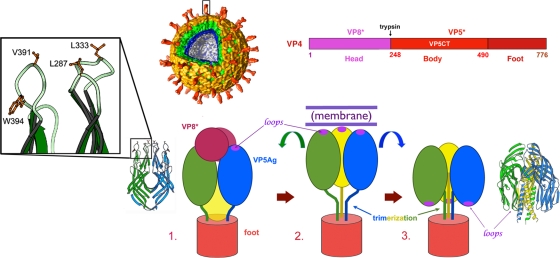
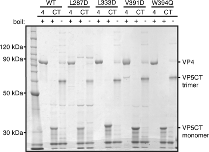
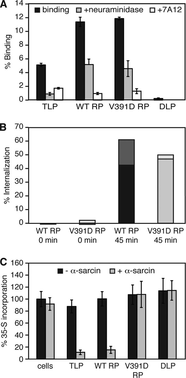
Experiments in cell-free systems have demonstrated that the VP5 cleavage fragment of the rotavirus spike protein, VP4, undergoes a foldback rearrangement that translocates three clustered hydrophobic loops from one end of the molecule to the other. This conformational change resembles the foldback rearrangements of enveloped virus fusion proteins. By recoating rotavirus subviral particles with recombinant VP4 and VP7, we tested the effects on cell entry of substituting hydrophilic for hydrophobic residues in the clustered VP5 loops. Several of these mutations decreased the infectivity of recoated particles without preventing either recoating or folding back. In particular, the V391D mutant had a diminished capacity to interact with liposomes when triggered to fold back by serial protease digestion in solution, and particles recoated with this mutant VP4 were 10,000-fold less infectious than particles recoated with wild-type VP4. Particles with V391D mutant VP4 attached normally to cells and internalized efficiently, but they failed in the permeabilization step that allows coentry of the toxin alpha-sarcin. These findings indicate that the hydrophobicity of the VP5 apex is required for membrane disruption during rotavirus cell entry.


Figures

FIG. 1.
FIG. 1.
Structures and model for conformational rearrangements of VP4. (Top center) Surface rendering from electron cryomicroscopy of a three-dimensional reconstruction of the rotavirus particle. A trypsin-cleaved VP4 spike (red) is boxed. The cutaway shows the multiple layers of the TLP. The VP7 layer is in yellow. The layers of the DLP are in green (VP6) and blue (VP2). (Top right) The VP4 primary structure indicating the boundaries of proteolytic products. (Bottom) Model for VP4 conformational rearrangements accompanying membrane penetration. (Step 1) Trypsin-activated VP4, in a schematic representation of a spike in roughly the orientation of the boxed spike in the rendering of a virion. The VP4 trimer has a 3-fold-symmetric “foot” but an asymmetrically organized projection. The ribbon diagram shows a dimeric form of the VP5 β-barrel domain (or antigen domain), which fits the dimer-clustered “body” of the projection, and the inset shows details of the three conserved hydrophobic loops that cap the β-barrel domain of VP5*. The hydrophobic residues mutated in this study are labeled. (Step 2) Dissociation of VP8* exposes the hydrophobic loops (shown as purple ovals) of VP5*. VP5* extends and engages a target membrane with the hydrophobic loops, probably from all three subunits. (Step 3) VP5* folds back to a stable trimeric structure, represented by the VP5CT crystal structure. This foldback is proposed to drive membrane penetration.
FIG. 2.
FIG. 2.
VP5CT formation by VP4 mutants. VP4 mutants serially digested with chymotrypsin and trypsin (CT) were mixed with reducing SDS-PAGE sample buffer and either heated to 95°C (+) or incubated at RT (−) before separation by SDS-PAGE and Coomassie staining. Purified VP4 (1 μg), heated and reduced, was loaded in lanes 4. A protein molecular mass ladder was loaded in the left lane.
FIG. 3.
FIG. 3.
Liposome interaction of VP4 loop mutants. (A) Liposome interaction of VP4 mutants serially digested with chymotrypsin and trypsin. (Left) Schematic of the digestion reaction showing the starting reactants and the putative liposome-interacting product. (Right) VP4 was serially digested in the presence of liposomes, and the mixtures were separated on discontinuous sucrose gradients (29). Samples of gradient fractions from top (lane 1) to bottom (lane 7) were not heated before separation by SDS-PAGE and immunoblotting with MAb 4D8 to detect VP5CT. (B) Liposome interaction of V391D VP5*. (Left) Schematic of the uncoating reaction showing the starting reactants and the putative liposome-interacting product. (Right) Purified RPs were uncoated in the presence of liposomes by adding 1 mM EDTA, and the mixtures were separated over discontinuous sucrose gradients. Samples of gradient fractions from top (lane 1) to bottom (lane 7) were heated prior to separation by SDS-PAGE and immunoblotting with MAb HS2 to detect VP5*.
FIG. 4.
FIG. 4.
VP4 incorporation into and infectivity of particles recoated with loop mutants of VP4. (A to C) Trypsin cleavage of assembled VP4 on RPs containing VP4V391D (A), VP4W394Q (B), and VP4L333D (C). Before separation of equivalent volumes by SDS-PAGE, the recoating products were digested with 5 μg/ml trypsin in 1 mM calcium (+T) or 5 mM EDTA (to uncoat the particles) (E/T) or were not digested but were diluted 1:50 (1:50 dil). VP4 and VP5* bands were detected by immunoblotting with MAb HS2. No VP4 or VP5* bands were detected after the VP4L333D recoating reaction products were incubated with EDTA and digested with trypsin (not shown). In panels C and F, VP4L333D concentrations (mg/ml) during VP4 recoating are indicated. (D to F) Infectivities of particles recoated with VP4V391D (D), VP4W394Q (E), and VP4L333D (F) and then primed with trypsin. Infectivity is reported relative to that of DLPs. In panel F, the recoating VP4WT concentrations at which VP4WT occupancy was equivalent to VP4L333D occupancy are indicated by an asterisk above each bar. The error bars represent standard deviations.
FIG. 5.
FIG. 5.
VP4V391D RP binding, internalization, and membrane permeabilization during cell entry. (A) VP4V391D RP binding to cells. 35S-labeled TLPs or RPs (∼5 × 10⁴ particles/cell) were bound to confluent MA104 cell monolayers for 1 h at 4°C. After washing and cell lysis in 1% SDS, the amount of bound virus was quantitated by liquid scintillation. Cells were pretreated with 200 mU/ml of V. cholerae neuraminidase for 1 h at 37°C before the addition of labeled virus (+neuraminidase), or particles were preincubated with MAb 7A12 (1 mg/ml) for 1 h at 37°C before being added to the cells (+7A12). The radioactivity detected from input particles was set as 100%. The error bars represent standard deviations. (B) Internalization of VP4V391D RPs. 35S-labeled VP4WT RPs or VP4V391D RPs (∼5 × 10⁴ particles/cell) were incubated with MA104 cell monolayers for 1 h at 4°C and washed. Some monolayers were then digested with 0.5 mg/ml proteinase K (0 min); others were incubated at 37°C for an additional 45 min before proteinase K digestion (45 min). The cells were lysed in 1% Triton X-100, and the associated label was quantitated by liquid scintillation. At each time point, the signal from parallel samples not digested with proteinase K was set as 100%. Values for duplicate trials are indicated by the dual shading of each bar. (C) α-Sarcin coentry. TLPs, VP4WT RPs, VP4V391D RPs, or DLPs (∼5 × 10⁴ particles/cell) or no particles, with or without α-sarcin (100 μg/ml), were incubated with confluent MA104 cell monolayers for 1 h at 37°C. Inocula were removed, and the cells were washed and then incubated in medium containing 35S-labeled Cys/Met (1 μCi/ml) for 1 h at 37°C. Following washing and TCA precipitation, radiolabel incorporation was quantitated by liquid scintillation counting. The amount of label incorporation in cells that were not exposed to α-sarcin or particles was set as 100%. The error bars represent standard deviations.

Similar articles

Cited by

References

    1. Agosto, M. A., T. Ivanovic, and M. L. Nibert. 2006. Mammalian reovirus, a nonfusogenic nonenveloped virus, forms size-selective pores in a model membrane. Proc. Natl. Acad. Sci. U. S. A. 103:16496-16501. - PMC - PubMed
    1. Aoki, S. T., E. C. Settembre, S. D. Trask, H. B. Greenberg, S. C. Harrison, and P. R. Dormitzer. 2009. Structure of rotavirus outer-layer protein VP7 bound with a neutralizing Fab. Science 324:1444-1447. - PMC - PubMed
    1. Bong, D. T., C. Steinem, A. Janshoff, J. E. Johnson, and M. Reza Ghadiri. 1999. A highly membrane-active peptide in Flock House virus: implications for the mechanism of nodavirus infection. Chem. Biol. 6:473-481. - PubMed
    1. Carrasco, L. 1995. Modification of membrane permeability by animal viruses. Adv. Virus Res. 45:61-112. - PMC - PubMed
    1. Chemello, M. E., O. C. Aristimuno, F. Michelangeli, and M. C. Ruiz. 2002. Requirement for vacuolar H+-ATPase activity and Ca2+ gradient during entry of rotavirus into MA104 cells. J. Virol. 76:13083-13087. - PMC - PubMed

Publication types

Substances

LinkOut - more resources