. 2010 Jun;151(6):2433-42.
doi: 10.1210/en.2009-1225. Epub 2010 Apr 7.

Progesterone is essential for maintenance and growth of uterine leiomyoma

Affiliations

Progesterone is essential for maintenance and growth of uterine leiomyoma

Hiroshi Ishikawa et al. Endocrinology. 2010 Jun.

Abstract

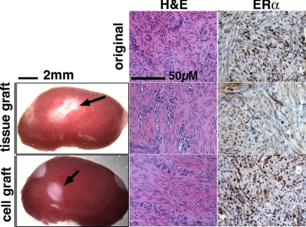
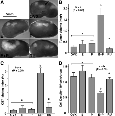
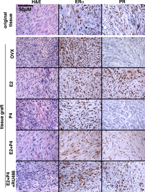
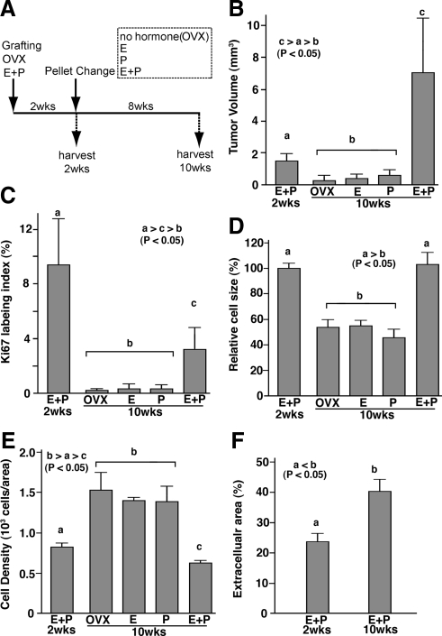
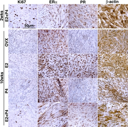
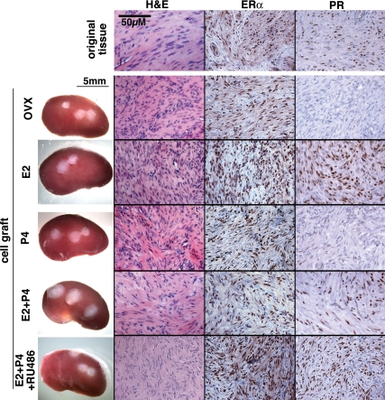
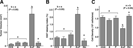
Uterine leiomyomata (ULs) represent the most common tumor in women and can cause abnormal uterine bleeding, large pelvic masses, and recurrent pregnancy loss. Although the dependency of UL growth on ovarian steroids is well established, the relative contributions of 17beta-estradiol and progesterone are yet to be clarified. Conventionally, estradiol has been considered the primary stimulus for UL growth, and studies with cell culture and animal models support this concept. In contrast, no research model has clearly demonstrated a requirement of progesterone in UL growth despite accumulating clinical evidence for the essential role of progesterone in this tumor. To elucidate the functions of ovarian steroids in UL, we established a xenograft model reflecting characteristics of these tumors by grafting human UL tissue beneath the renal capsule of immunodeficient mice. Leiomyoma xenografts increased in size in response to estradiol plus progesterone through cell proliferation and volume increase in cellular and extracellular components. The xenograft growth induced by estradiol plus progesterone was blocked by the antiprogestin RU486. Furthermore, the volume of established UL xenografts decreased significantly after progesterone withdrawal. Surprisingly, treatment with estradiol alone neither increased nor maintained the tumor size. Although not mitogenic by itself, estradiol induced expression of progesterone receptor and supported progesterone action on leiomyoma xenografts. Taken together, our findings define that volume maintenance and growth of human UL are progesterone dependent.


Figures

Figure 1
Figure 1
Gross appearance and histology of myometrial xenografts. Gross appearance of myometrial tissue and cell xenografts (indicated by an arrow) on the host kidney (treated with E2+P4) at 8 wk after grafting. Although the myometrial xenografts did not increase in size with any hormone treatment, both tissue and cell xenografts (with E2 treatment) showed typical histology of myometrium comparable with the original tissue (top panels) in H&E and ERα-IHC staining.
Figure 2
Figure 2
Effect of E2 and P4 on the growth of UL xenografts. OVX, OVX control (no hormone); E, E2 treated; P, P4 treated; E+P, E2+P4 treated; RU, E2+P4+RU486 treated groups. UL tissues were grown as subrenal grafts in OVX mouse host treated with E2 (E), P4 (P), E2+P4 (E+P), or E2+P4+RU486 (RU) for 8 wk. Gross appearance of xenografts on host kidney (A; graft is indicated by an arrow), tumor volume (B), Ki67 labeling index (C), and cell density (D). The bar indicates average value ± sem. Statistical differences were assessed by using the Kruskal-Wallis test (n = 6, P < 0.05). UL tissues enlarged in response to E2 plus P4 treatment (A and B). E2+P4 also increased ki67 labeling index (C) and decreased cell density (D). This growth-promoting effect of E2 plus P4 treatment was blocked by RU486. E2 and P4 alone had no effect on the growth of UL tissues.
Figure 3
Figure 3
Histology of UL xenografts and original tumor. OVX, No hormone control. UL xenografts survived for 8 wk and retained histology typical for leiomyoma in all hormone treatment groups. Whereas ERα was expressed in all xenografts irrespective of hormone treatment, expression of PR was dependent on E2. Thus, PR was undetectable in OVX and P4 groups. All histology images were taken under a ×20 objective lens and in the same magnification.
Figure 4
Figure 4
Effect of hormone withdrawal on UL xenograft. A, Time line. UL xenografts were grown for 2 wk with E2+P4 (E+P 2wks), and then hormone pellets were replaced with E2+P4 (E+P), E2 (E), P4 (P), or no hormone (OVX). Eight weeks after pellet replacement (10 wk after grafting), xenografts were harvested and analyzed. Tumor volume (B), Ki67 labeling index (C), relative cell size (D; relative value to average cell size at 2 wk after grafting), cell density (E), and extracellular area (β-actin negative area; F). Bars indicate average value ± sem. Statistical differences were assessed by using the Kruskal-Wallis test (n = 6, P < 0.05). Tumor volume increased only with E2+P4 treatment (B). In response to withdrawal of E2 and/or P4, tumor volume (B), Ki67 labeling index (C), and cell size (D) significantly declined. Accordingly, cell density increased in OVX, E, and P groups (E). In the E+P group, cell size remained the same; however, the cell density decreased significantly (E) with increase of extracellular component (F) from 2 to 10 wk after grafting, indicating tumor volume increased by both cell number and ECM volume.
Figure 5
Figure 5
Histology of UL xenografts subjected to hormone withdrawal. Proliferation activity significantly declined in response to withdrawal of E2 and/or P4 as assessed by Ki67 expression. PR was also down-regulated in response to E2 withdrawal. However, UL xenografts did not show sign of tissue degradation or cell death, and expression of ERα and β-actin was maintained without E2 and/or P4, even after 8 wk. All histology images were taken under a ×20 objective lens and in the same magnification.
Figure 6
Figure 6
Gross appearance and the histology of UL cell xenografts. Xenografts constructed with 106 dissociated leiomyoma cells formed tumors with typical histology of UL. Expression of ERα and PR was also comparable with that of tissue xenografts (Fig. 3). All histology images were taken under a ×20 objective lens and in the same magnification.
Figure 7
Figure 7
Growth of UL cell xenografts. OVX, OVX control (no hormone); E, E2 treated; P, P4 treated; E+P, E2+P4 treated; RU, E2+P4+RU486 treated groups. UL cell xenografts were grown in OVX mouse host treated with E2 (E), P4 (P), E2+P4 (E+P), or E2+P4+RU486 (RU) for 8 wk. As assessed by tumor volume (A), Ki67 labeling index (B), and cell density (C), the response of UL cell xenografts to E2 and P4 was essentially identical with that of tissue xenografts (Fig. 2). Xenografts increased their size in response to E2 plus P4 treatment (A). E2+P4 also increased ki67 labeling index (B) and decreased cell density (C). This growth-promoting effect of E2 plus P4 treatment was blocked by RU486. E2, P4 alone had no effect on the growth of UL cell grafts. The bars indicate average value ± sem. Statistical differences were assessed by using the Kruskal-Wallis test (n = 6, P < 0.05).

Similar articles

Cited by

References

    1. Day Baird D, Dunson DB, Hill MC, Cousins D, Schectman JM 2003 High cumulative incidence of uterine leiomyoma in black and white women: ultrasound evidence. Am J Obstet Gynecol 188:100–107 - PubMed
    1. Parker WH 2007 Etiology, symptomatology, and diagnosis of uterine myomas. Fertil Steril 87:725–736 - PubMed
    1. Merrill RM 2008 Hysterectomy surveillance in the United States, 1997 through 2005. Med Sci Monit 14:CR24–CR31 - PubMed
    1. Shimomura Y, Matsuo H, Samoto T, Maruo T 1998 Up-regulation by progesterone of proliferating cell nuclear antigen and epidermal growth factor expression in human uterine leiomyoma. J Clin Endocrinol Metab 83:2192–2198 - PubMed
    1. Park SH, Ramachandran S, Kwon SH, Cha SD, Seo EW, Bae I, Cho C, Song DK 2008 Upregulation of ATP-sensitive potassium channels for estrogen-mediated cell proliferation in human uterine leiomyoma cells. Gynecol Endocrinol 24:250–256 - PubMed

Publication types