Comparative Study
. 2010 Jun;48(6):2116-21.
doi: 10.1128/JCM.02530-09. Epub 2010 Apr 7.

Development of a loop-mediated isothermal amplification assay for rapid detection of subgroup J avian leukosis virus

Affiliations
Comparative Study

Development of a loop-mediated isothermal amplification assay for rapid detection of subgroup J avian leukosis virus

Xiaotao Zhang et al. J Clin Microbiol. 2010 Jun.

Abstract

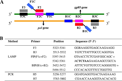
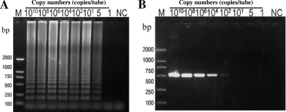
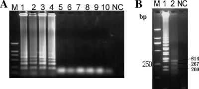
Infection of breeder flocks in China with subgroup J avian leukosis virus (ALV-J) has increased recently. In this study, we have developed a loop-mediated isothermal amplification (LAMP) assay for rapid detection of ALV-J from culture isolates and clinical samples. The ALV-J-specific LAMP assay efficiently amplified the target gene within 45 min at 63 degrees C using only a simple laboratory water bath. To determine the specificity of the LAMP assay, various subgroup ALVs and other related viruses were detected. A ladder pattern on gel electrophoresis was observed for ALV-J isolates but not for other viruses. To evaluate the sensitivities of the LAMP assay and conventional PCR, the NX0101 isolate plasmid DNA was amplified by them. The detection limit of the LAMP assay was 5 target gene copies/reaction, which was up to 20 times higher than that of conventional PCR. To evaluate the application of the LAMP assay for detection of ALV-J in clinical samples, 49 samples suspected of ALV infection from breeder flocks were tested by the LAMP assay and PCR. Moreover, virus isolation from these samples was also performed using cell culture. The positive-sample ratios were 21/49 (43%) by conventional PCR, 26/49 (53%) by the LAMP assay, and 19/46 (41%) by virus isolation. Additionally, a positive LAMP reaction can be visually ascertained by the observation of turbidity or a color change after addition of SYBR green I dye. Consequently, the LAMP assay is a simple, rapid, and sensitive diagnostic method and can potentially be developed for rapid detection of ALV-J infection in the field.


Figures

FIG. 1.
FIG. 1.
Loop-mediated isothermal amplification (LAMP) and PCR primer design for detection of ALV-J. (A) Locations of LAMP primers in relation to the defined regions of the target sequence. The forward outer primer (F3) and the forward inner primer (FIP) are complementary to the indicated region of the pol gene (arrows). The reverse outer primer (B3) and the reverse inner primer (BIP) are complementary to the indicated region of the gp85 gene (arrows). FIP and BIP contain two distinct sequences (F1c plus F2 and B1c plus B2, respectively). (B) The LAMP and PCR primer positions are shown relative to prototype ALV-J clone HPRS-103 (GenBank accession number Z46390). The DdeI restriction site is indicated in bold type (R = G/A and Y = C/T).
FIG. 2.
FIG. 2.
Comparative sensitivities of the LAMP assay and PCR for detection of ALV-J by agarose gel electrophoresis. The plasmid containing the NX0101 gene fragment was serially diluted. Each plasmid concentration was subjected to the LAMP assay and conventional PCR. (A) ALV-J-specific LAMP reaction. (B) PCR revealing a 545-bp specific amplicon for ALV-J. Lane M, DNA marker DL-2000 (TaKaRa, Japan); NC, negative control.
FIG. 3.
FIG. 3.
Specificity of the LAMP assay. (A) Agarose gel electrophoresis analysis of different subgroup ALVs. Lanes 1 through 8, LAMP of DNA from the Js-nt, SCAU-0901, SCAU-CHN, NX0101, GD08, RAV-1, RAV-2, and CD08 isolates, respectively; lanes 9 through 10, LAMP of DNA from subgroup E (E1 and E2). (B) Restriction enzyme analysis of LAMP products on a 3% agarose gel. Lane 1, undigested LAMP product of the NX0101 isolate; lane 2, LAMP product after digestion with DdeI. Lane M, DNA marker DL-2000 (TaKaRa, Japan); NC, negative control.
FIG. 4.
FIG. 4.
Visual detection of LAMP products. (A) Turbidity observation. Turbidity increases in the positive reaction due to the generation of magnesium pyrophosphate. (B) ALV-J LAMP products were detected after addition of SYBR green I dye. The positive reaction shows a color change to yellowish green, which can be easily distinguished from the reddish orange color indicating a negative reaction.

Similar articles

Cited by

References

    1. Bai, J., L. N. Payne, and M. A. Skinner. 1995. HPRS-103 (exogenous avian leukosis virus, subgroup J) has an env gene related to those of endogenous elements EAV-0 and E51 and an E element found previously only in sarcoma viruses. J. Virol. 69:779-784. - PMC - PubMed
    1. Benson, S. J., B. L. Ruis, A. L. Garbers, A. M. Fadly, and K. F. Conklin. 1998. Independent isolates of the emerging subgroup J avian leukosis virus derive from a common ancestor. J. Virol. 72:10301-10304. - PMC - PubMed
    1. Bista, B. R., C. Ishwad, A. Vats, P. Manna, G. Gupta, M. Adhikari, et al. 2007. Development of a loop-mediated isothermal amplification assay for rapid detection of BK virus. J. Clin. Microbiol. 45:1581-1587. - PMC - PubMed
    1. Bova, C. A., J. P. Manfredi, and R. Swanstrom. 1986. Env genes of avian retroviruses: nucleotide sequence and molecular recombinants define host range determinants. Virology 152:343-354. - PubMed
    1. Bova, C. A., J. C. Olsen, and R. Swanstrom. 1988. The avian retrovirus env gene family: molecular analysis of host range and antigenic variants. J. Virol. 62:75-83. - PMC - PubMed

Publication types

LinkOut - more resources