. 2010 Jul;299(1):C33-41.
doi: 10.1152/ajpcell.00033.2010. Epub 2010 Apr 7.

Deletion of the anion exchanger Slc26a4 (pendrin) decreases apical Cl(-)/HCO3(-) exchanger activity and impairs bicarbonate secretion in kidney collecting duct

Affiliations

Deletion of the anion exchanger Slc26a4 (pendrin) decreases apical Cl(-)/HCO3(-) exchanger activity and impairs bicarbonate secretion in kidney collecting duct

Hassane Amlal et al. Am J Physiol Cell Physiol. 2010 Jul.

Abstract

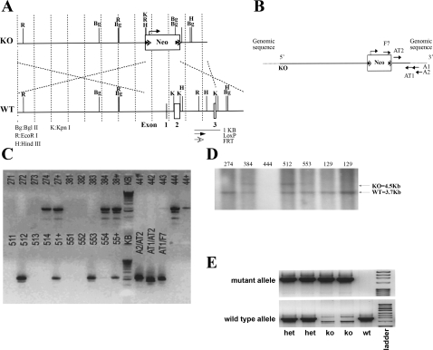
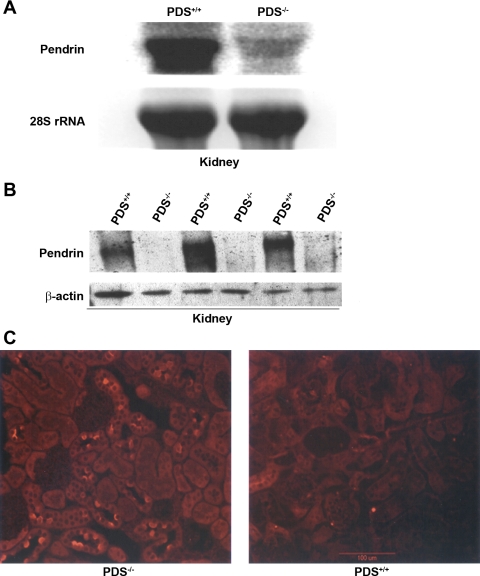
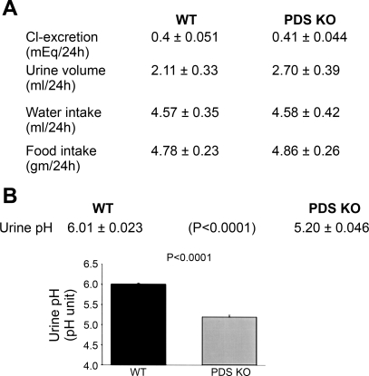
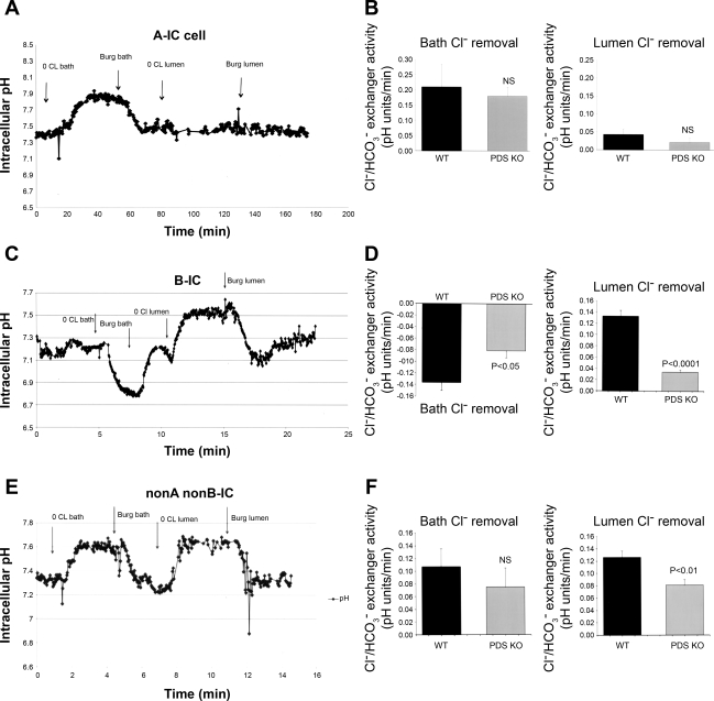
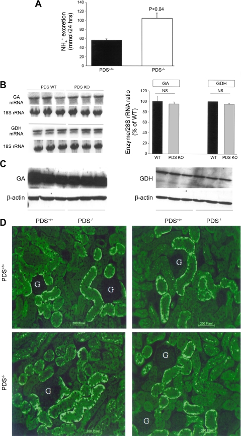
The anion exchanger Pendrin, which is encoded by SLC26A4 (human)/Slc26a4 (mouse) gene, is localized on the apical membrane of non-acid-secreting intercalated (IC) cells in the kidney cortical collecting duct (CCD). To examine its role in the mediation of bicarbonate secretion in vivo and the apical Cl(-)/HCO(3)(-) exchanger in the kidney CCD, mice with genetic deletion of pendrin were generated. The mutant mice show the complete absence of pendrin expression in their kidneys as assessed by Northern blot hybridization, Western blot, and immunofluorescence labeling. Pendrin knockout (KO) mice display significantly acidic urine at baseline [pH 5.20 in KO vs. 6.01 in wild type (WT); P < 0.0001] along with elevated serum HCO(3)(-) concentration (27.4 vs. 24 meq/l in KO vs. WT, respectively; P < 0.02), consistent with decreased bicarbonate secretion in vivo. The urine chloride excretion was comparable in WT and KO mice. For functional studies, CCDs were microperfused and IC cells were identified by their ability to trap the pH fluorescent dye BCECF. The apical Cl(-)/HCO(3)(-) exchanger activity in B-IC and non-A, non-B-IC cells, as assessed by intracellular pH monitoring, was significantly reduced in pendrin-null mice. The basolateral Cl(-)/HCO(3)(-) exchanger activity in A-IC cells and in non-A, non-B-IC cells, was not different in pendrin KO mice relative to WT animals. Urine NH(4)(+) (ammonium) excretion increased significantly, consistent with increased trapping of NH(3) in the collecting duct in pendrin KO mice. We conclude that Slc26a4 (pendrin) deletion impairs the secretion of bicarbonate in vivo and reduces apical Cl(-)/HCO(3)(-) exchanger activity in B-IC and non-A, non-B-IC cells in CCD. Additional apical Cl(-)/HCO(3)(-) exchanger(s) is (are) present in the CCD.


Figures

Fig. 1.
Fig. 1.
Generation of Slc26a4−/− mutant mice. A: schematic diagram of the Slc26a4 targeting construct. The strategy was to delete the Slc26a4 gene by knocking out exons 1–3, which encode 101 amino acids including the ATG initiation site in exon 2. The construct was designed with the short homology arm (SA) extending 1.6 kb 3′ to exon 3 and the long homology arm (LA) located on the 5′ side of exon 1, with ∼7.6-kb length. The Neocassette (Neo) replaces 4.2 kb of the gene including exons 1–3. The targeting vector was confirmed by restriction analysis after each modification step and by sequencing using primers designed to read from the selection cassette into the 3′ end of the SA (N1) and the 5′ end of the LA (N2) or primers that anneal to the vector sequence, P6 and T7, and read into the 5′ and 3′ ends of the bacterial artificial chromosome (BAC) subclone. KO, knockout; WT, wild type. B: Slc26a4 targeting allele and delineation of locations of primers. Ten micrograms of the targeting vector was linearized by NotI and then transfected by electroporation of C57BL/6 × 129/SvEv hybrid embryonic stem cells (ES). After selection in G418 antibiotic, surviving clones were expanded for PCR analysis to identify recombinant ES clones. Primers A1 and A2 were designed downstream (3′) to the SA outside the region used to generate the targeting construct. C: identification of homologous recombinant clones. PCR reactions using A1 and A2 with the F7 primer at the 3′ end of the Neocassette amplify 1.6- and 1.9-kb fragments, respectively. The control PCR reaction was done with primers AT1 and AT2, which are at the 3′ and 5′ ends of the SA, respectively, inside the region used to create the targeting construct. This amplifies a band of 1.5 kb. PCR analysis of DNA isolated from >100 surviving colonies identified several individual clones that showed homologous recombination as determined by the presence of a 1.9-kb fragment. The positive control was performed with primers AT1/N1, which gave the expected fragment size of 1.9 kb. D: Southern blot analysis. For Southern blotting, the fragment encoded by EcoR1-EcoRI, which spans from the 5′ end of the Neocassette to the 3′ end of the SA (A) was amplified for verification of homologous recombination. Two ES clones, 384 and 512, that were identified by PCR screening (C) were further examined by Southern blotting and showed homologous recombination as shown by the presence of a 4.5-kb fragment. E: generation of Slc26a4+/+ and Slc26a4−/− mice. Tail DNA genotyping identified Slc26a7+/+ (wt), +/− (het), and −/− (ko) mice. Band at top shows the detection of the mutant allele, and band at bottom represents the WT allele.
Fig. 2.
Fig. 2.
Generation of Slc26a4−/− mutant mice. A: expression of Slc26a4 in the kidney. Crossing of male and female heterozygote mice (+/−) resulted in the generation of Slc26a4 KO (−/−) mice. Northern blot hybridization on RNA isolated from kidneys of Slc26a4 +/+ and −/− mice indicated that the expression of Slc26a4 is completely absent in the Slc26a4-null mouse. The faint larger band in KO mice represents the mutant RNA. PDS, pendrin. B: immunoblot analysis of pendrin. Microsomal membrane proteins from kidneys of Slc26a4+/+ and Slc26a4−/− mice were isolated, resolved on a gel, and blotted against pendrin antibody. Bottom: β-actin expression as a control for loading. Top: results demonstrate the complete ablation of pendrin in kidneys of KO mice. C: immunofluorescence labeling. Immunofluorescence labeling was performed on frozen kidney sections as previously described (18). As noted, the expression of pendrin, which is located on the apical membrane of a subset of cortical collecting duct (CCD) cells (right) was completely abolished in pendrin KO mice (left).
Fig. 3.
Fig. 3.
Balanced studies in pendrin KO and WT mice. A: balanced studies and urine chemical analysis in WT and mutant mice. Animals were placed in metabolic cages and allowed free access to food and water for 72 h before the experiments started. Urine output, water intake, food intake, and urine chloride concentration were measured. The 24-h urine chloride as well as urine volume and water intake are not different in pendrin-null mice relative to WT animals. B: urine pH measurements in pendrin KO and WT mice. Urine pH was measured with a pH-sensitive microelectrode from samples collected in animals placed in metabolic cages. As shown, urine pH was significantly decreased in pendrin KO mice.
Fig. 4.
Fig. 4.
Intracellular pH (pHi) monitoring in intercalated (IC) cells in microperfused CCD. A: representative pHi tracings in A-IC cells in Slc26a4+/+ mouse. CCDs of WT or KO animals were microperfused in vitro. A-IC cells were identified by the presence of Cl/HCO3 exchange limited to their basolateral membrane. Removal of bath chloride causes intracellular alkalinization, whereas the removal of luminal chloride has no significant effect on pHi. Burg solution refers to the chloride-containing solution originally used in Dr. Burg's laboratory (see experimental proceduresand Ref. 3). B: pHi summary results in A-IC cells. There was no significant difference in WT and pendrin KO mice. The results of experiments comparing the basolateral Cl/HCO3 exchanger activity did not show any significant difference in WT and KO mice. NS, not significant. C: representative pHi tracings in B-IC cells in Slc26a4+/+ mouse. CCDs of WT or KO animals were microperfused in vitro. B-IC cells were identified by the presence of Cl/HCO3 exchange limited to their apical membrane. Removal of luminal chloride causes intracellular alkalinization, whereas the removal of bath chloride causes acidification. D: pHi summary results in B-IC cells. The results of experiments comparing the apical Cl/HCO3 exchanger activity showed significant reduction in B-IC cells in pendrin KO mice. E: representative pHi tracings in non-A, non-B-IC cells in Slc26a4+/+ mouse. CCD of WT or KO animals were microperfused in vitro. Non-A, non-B-IC cells were identified by the presence of Cl/HCO3 exchange to both apical and basolateral membranes. Removal of bath or luminal chloride causes intracellular alkalinization. F: pHi summary results in non-A, non-B-IC cells. The results of experiments comparing the apical Cl/HCO3 exchanger activity showed significant reduction in non-A, non-B-IC cells in pendrin KO mice. The basolateral Cl/HCO3 exchanger activity was not significantly different in non-A, non-B-IC cells in pendrin KO mice.
Fig. 5.
Fig. 5.
Renal NH4+ excretion in pendrin KO and WT mice. A: 24-h urine NH4+ excretion. Twenty-four-hour urinary excretion of NH4+ was significantly increased in pendrin KO mice. B: expression of glutamine dehydrogenase (GDH) and phosphate-dependent glutaminase (GA). Left: mRNA expression of GDH and GA. Right: quantitation of GDH and GA mRNA expression levels in WT and KO mice. C: protein abundance of GDH and phosphate-dependent GA. Western blot analysis demonstrated that the protein abundance of GDH and GA remained unchanged in pendrin KO mice relative to WT littermates. D: immunofluorescence labeling of the Na+-HCO3 cotransporter NBC1. Expression of kNBC1 in the kidney proximal tubule was examined by immunofluorescence labeling in pendrin KO mice (bottom) and WT littermates (top). As shown, NBC1 expression did not show any significant difference in the kidneys of pendrin KO animals relative to WT mice. G, glomerulus.

Similar articles

Cited by

References

    1. Alper SL, Natale J, Gluck S, Lodish HF, Brown D. Subtypes of intercalated cells in rat kidney collecting duct defined by antibodies against erythroid band 3 and renal vacuolar H+-ATPase. Proc Natl Acad Sci USA 86: 5429–5433, 1989 - PMC - PubMed
    1. Amlal H, Sheriff S, Faroqui S, Ma L, Barone S, Petrovic S, Soleimani M. Regulation of acid-base transporters by vasopressin in the kidney collecting duct of Brattleboro rat. Am J Nephrol 26: 194–205, 2006 - PubMed
    1. Atkins JL, Burg MB. Bicarbonate transport by isolated perfused rat collecting ducts. Am J Physiol Renal Fluid Electrolyte Physiol 249: F485–F489, 1985 - PubMed
    1. Bissig M, Hagenbuch B, Stieger B, Koller T, Meier PJ. Functional expression cloning of the canalicular sulfate transport system of rat hepatocytes. J Biol Chem 269: 3017–3021, 1994 - PubMed
    1. Brown D, Breton S. H+V-ATPase-dependent luminal acidification in the kidney collecting duct and the epididymis/vas deferens: vesicle recycling and transcytotic pathways. J Exp Biol 203: 137–145, 2000 - PubMed

Publication types

MeSH terms

LinkOut - more resources