. 2010 Apr 27;107(17):7957-62.
doi: 10.1073/pnas.0912478107. Epub 2010 Apr 7.

Temporal delays among place cells determine the frequency of population theta oscillations in the hippocampus

Affiliations

Temporal delays among place cells determine the frequency of population theta oscillations in the hippocampus

Caroline Geisler et al. Proc Natl Acad Sci U S A. .

Abstract

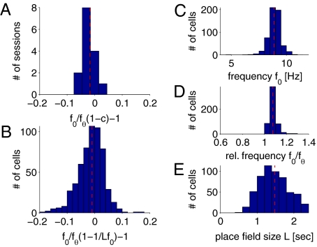
Driven either by external landmarks or by internal dynamics, hippocampal neurons form sequences of cell assemblies. The coordinated firing of these active cells is organized by the prominent "theta" oscillations in the local field potential (LFP): place cells discharge at progressively earlier theta phases as the rat crosses the respective place field ("phase precession"). The faster oscillation frequency of active neurons and the slower theta LFP, underlying phase precession, creates a paradox. How can faster oscillating neurons comprise a slower population oscillation, as reflected by the LFP? We built a mathematical model that allowed us to calculate the population activity analytically from experimentally derived parameters of the single neuron oscillation frequency, firing field size (duration), and the relationship between within-theta delays of place cell pairs and their distance representations ("compression"). The appropriate combination of these parameters generated a constant frequency population rhythm along the septo-temporal axis of the hippocampus, while allowing individual neurons to vary their oscillation frequency and field size. Our results suggest that the faster-than-theta oscillations of pyramidal cells are inherent and that phase precession is a result of the coordinated activity of temporally shifted cell assemblies, relative to the population activity, reflected by the LFP.


Conflict of interest statement

The authors declare no conflict of interest.

Figures

Fig. 1.
Fig. 1.
Place cells oscillate faster than the theta LFP. (A) Example place cells active in different parts of the maze. Different colors correspond to the action potentials of different neurons. (B) Spiking activity of a neuron (blue ticks) and LFP in a single run (1 s is shown). (C) Rat's position in the maze (distance from the delay area) vs. theta phase demonstrates the precession of spiking phase for two neurons (red and blue, from A). (D) Power spectra (normalized) of unit firing (blue, peak power frequency 9 Hz; red, peak power frequency 9.6 Hz) and corresponding LFP (gray dashed line, peak power frequency 7.9 Hz). Note that the units oscillate faster than the LFP. (E) Temporal cross-correlation between the two place cells (red and blue in A, C, and D). Dashed line, smoothed rate of neuronal spiking to eliminate theta modulation. Temporal distance T is the time needed for the rat to run the distance between the peaks of the two place fields (T = −295 ms). Inset: τ is the distance of the first theta peak from zero and corresponds to the distance between the two neurons within the theta cycle (τ = −30 ms). (F) Correlation between the peaks of place fields vs. theta-scale time lag τ for 3,352 pairs of neurons (data from ref. 20) is well fit by a sigmoid (red solid line) and by a linear fit in the central region (red dashed line; slope = 1.89 ms/cm). Top and Right: Histograms of distance and time lag, respectively.
Fig. 2.
Fig. 2.
Single-session examples of neuronal populations. (A) CA1 pyramidal cells during running on the maze while performing an alternation memory task. Far Left: Power spectra of simultaneously recorded place cells and the histogram of their oscillation frequencies. Black line, spectrum of the LFP (gray dashed line, peak power at 8.09 Hz); black dotted line, mean oscillation frequency of place cells (8.61 Hz). Middle Left: Histogram of oscillation frequencies, relative to the LFP (red dashed line, mean relative frequency f 0/fθ = 1.08 Hz). Middle Right: Theta time lag τ vs. travel time between place fields T. The slope of the linear regression fit (red dashed line) is the compression factor (c = 0.075). Far Right: Histogram of firing field sizes (durations) L. Mean place field duration is 1.5 s. (B) Simultaneously recorded wheel cells during wheel running from the same session as in A; LFP peak power at 7.32 Hz and mean place cell oscillation frequency is 7.71 Hz. Note that the oscillation frequency of both LFP and units is slower in the wheel than on the track.
Fig. 3.
Fig. 3.
Population of pyramidal cells oscillates at the same frequency as the LFP. (A) Raster plot of 23 simultaneously recorded CA1 pyramidal cells in a 3-s sample epoch. Most pyramidal cells did not fire spikes during the epoch. (B) Population output of place cells (POP) shown in A. (C) Frequency spectra of the POP in successive epochs. Red is high power, blue is low power. Black dots mark the location of the peak power of the LFP (see E). (D) Filtered LFP trace and (E) frequency spectra in the same epochs as in A–C. Black dots, peak power of LFP. (F) Spectra of three example neurons (peak power at 8.71 Hz, 8.56 Hz, and 9.02 Hz, respectively) and the average of the frequency spectra calculated separately for individual neurons (peak power at 8.56 Hz) (G). (H) Power spectra of the POP (red) and LFP (green), calculated from the selected epoch (peak power at 8.25 Hz). Note that whereas single pyramidal cells oscillate faster than the LFP theta, the oscillation frequency of the POP and the LFP are identical.
Fig. 4.
Fig. 4.
Model for generating population frequency slower than the oscillation frequencies of its constituents. Single neurons are characterized by the discharge probability (Methods) and phase shifted with respect to each other according to the compression factor c (parameters: single neuron oscillation frequency f 0 = 8.6 Hz, place field size L = 1.5 s, c = 0.075). (A) Three example neurons (color coded) with identical oscillation frequency but different phase offset, according to their maximal discharge location. Black dashed line, population output of model place cells (mPOP); see below. (B) A population of model neurons. The summed activity of the entire population (black dashed line) oscillates slower than each individual neuron (fθ = 8 Hz) with amplitude A. (C) The phase of the three example neurons (see A) with respect to the oscillation of the mPOP is plotted against time. Spikes are generated randomly from the single neuron discharge probability. Note that the neurons phase precess ≈360°. Right: Spike density for the example neurons.
Fig. 5.
Fig. 5.
Modeling the population output of model place cells (mPOP) on the basis of experimental data from Fig. 2. (A) Using the experimentally derived parameters during track running (mean frequency of single neurons f 0 = 8.61 Hz, compression factor c = 0.075, and firing field size L = 1.5 s), the oscillation frequency of the population output of the model place cells is fθ = 7.97 Hz (compare with the measured LFP frequency fθ = 8.09 Hz). (B) Histogram shows the distribution of single unit oscillation frequencies above the power spectrum of the population activity mPOP shown in A. Red dashed line, mean frequency of single neurons; black dashed line, peak frequency of mPOP. (C) Same as in A, using experimentally derived parameters from wheel running (f 0 = 7.71 Hz, c = 0.059, L = 2.15 s). (D) The oscillation frequency of the mPOP (fθ = 7.25 Hz, compare with the measured LFP frequency fθ = 7.32 Hz) is much lower than the mean frequency of single neurons (red dashed line).
Fig. 6.
Fig. 6.
Comparison between model predictions and experimental observations. (A) Histogram of the relative error between the relative average frequency predicted from the experimentally measured compression factor (1 − c) and the relative frequency f 0/fθ, averaged over the respective session. The mean error (red dashed line) is −0.017 (corresponding to >98% accuracy) for 16 recording sessions. (B) Histogram of the relative error between the relative frequency predicted from the place field size and the single neuron frequency [1 − 1/(Lf 0)], and the measured relative neuron frequency f 0/fθ for 680 pyramidal cells. The relative mean error (red dashed line) is −0.0097 (corresponding to >99% accuracy). (C) Histogram of single neuron frequencies f 0 (red dashed line, mean frequency 8.85 Hz), (D) relative frequencies f 0/fθ (red dashed line, mean relative frequency 1.07 Hz), and (E) place field size L (red dashed line, mean place field size 1.48 s) for 680 pyramidal cells.
Fig. 7.
Fig. 7.
Modeling the coherent theta frequency oscillations along the septo–temporal (dorsal–ventral) axis of the hippocampus. Pyramidal cells across the whole hippocampus oscillate at different frequencies, but their mPOP synchronizes at theta frequency. (A) The oscillation frequency f 0 decreases and the place field size L increases along the dorsal–ventral axis according to fθ = f 0 – 1/L, whereas the oscillation frequency of the mPOP does not change (fθ = 8 Hz). (B) Scheme of the hippocampus, color coded for decreasing oscillation frequencies of place cells, along the dorsal–ventral axis. (C) Firing fields of 20 selected place neurons, color coded to indicate their anatomical location in the dorsal–ventral axis (see B). (D) mPOP of 100 model pyramidal cells with randomly chosen oscillation frequencies (black line) and the analytically predicted 8 Hz population oscillation (gray line). (E) Enlargement of the traces of D. Note that the oscillation frequency of both lines is the same.

Similar articles

Cited by

References

    1. Friston KJ. The labile brain. I. Neuronal transients and nonlinear coupling. Philos Trans R Soc Lond B Biol Sci. 2000;355:215–236. - PMC - PubMed
    1. Logothetis NK. The underpinnings of the BOLD functional magnetic resonance imaging signal. J Neurosci. 2003;23:3963–3971. - PMC - PubMed
    1. Buzsáki G, Draguhn A. Neuronal oscillations in cortical networks. Science. 2004;304:1926–1929. - PubMed
    1. Steriade M, McCormick DA, Sejnowski TJ. Thalamocortical oscillations in the sleeping and aroused brain. Science. 1993;262:679–685. - PubMed
    1. Petsche H, Stumpf C, Gogolak G. [The significance of the rabbit's septum as a relay station between the midbrain and the hippocampus. I. The control of hippo-campus arousal activity by the septum cells] Electroencephalogr Clin Neurophysiol. 1962;14:202–211. - PubMed

Publication types

LinkOut - more resources