Comparative Study
. 2010 May 15;184(10):5466-5474.
doi: 10.4049/jimmunol.0903561. Epub 2010 Apr 7.

B7-H1 expression on old CD8+ T cells negatively regulates the activation of immune responses in aged animals

Affiliations
Comparative Study

B7-H1 expression on old CD8+ T cells negatively regulates the activation of immune responses in aged animals

Noweeda Mirza et al. J Immunol. .

Abstract

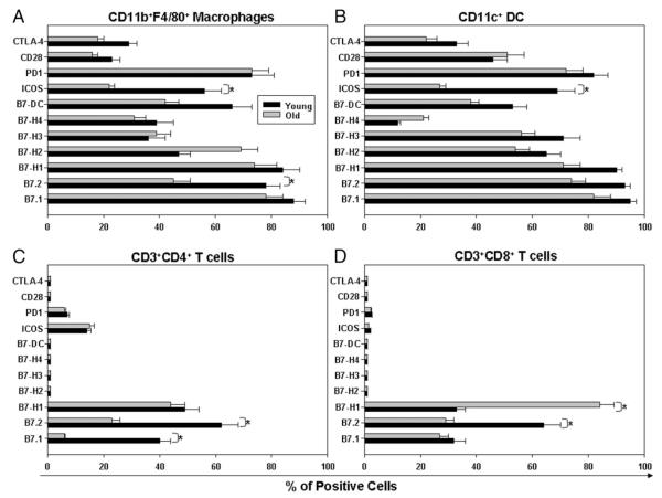
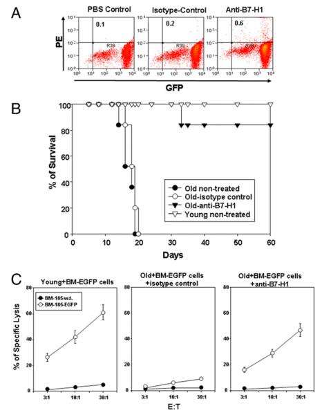
T cell responses are compromised in the elderly. The B7-CD28 family receptors are critical in the regulation of immune responses. We evaluated whether the B7-family and CD28-family receptors were differentially expressed in dendritic cells, macrophages, and CD4(+) and CD8(+) T cells from young and old mice, which could contribute to the immune dysfunction in the old. Although most of the receptors were equally expressed in all cells, >85% of the old naive CD8(+) T cells expressed B7-H1 compared with 25% in the young. Considering that B7-H1 negatively regulates immune responses, we hypothesized that expression of B7-H1 would downregulate the function of old CD8(+) T cells. Old CD8(+) T cells showed reduced ability to proliferate, but blockade of B7-H1 restored the proliferative capacity of old CD8(+) T cells to a level similar to young CD8(+) T cells. In vivo blockade of B7-H1 restored antitumor responses against the B7-H1(-) BM-185-enhanced GFP tumor, such that old animals responded with the same efficiency as young mice. Our data also indicate that old CD8(+) T cells express lower levels of TCR compared with young CD8(+) T cells. However, following antigenic stimulation in the presence of B7-H1 blockade, the levels of TCR expression were restored in old CD8(+) T cells, which correlated with stronger T cell activation. These studies demonstrated that expression of B7-H1 in old CD8(+) T cells impairs the proper activation of these cells and that blockade of B7-H1 could be critical to optimally stimulate a CD8 T cell response in the old.


Figures

FIGURE 1
FIGURE 1
Staining of young and old macrophages, DCs, and CD4 and CD8 T cells for the expression of B7 family and CD28 family receptors. A, Macrophages were stained for anti–CD11b-allophycocyanin and anti-F4/80-PE. B, DCs were stained with anti–CD11c-PE. C, CD4+ T cells were stained with anti–CD4-PE. D, CD8+ T cells were stained with anti–CD8-PE. The prevalence of B7 family and CD28 family receptors in these subpopulations was analyzed by staining the samples with FITC-conjugated Abs to anti-B7.1, anti-B7.2, anti–B7-H1, anti–B7-H2, anti–B7-H3, anti–B7-H4, anti–B7-DC, anti-ICOS, anti–PD-1, anti-CD28, and anti-CTLA4. The results shown are the average percentages of positive cells in the spleens from a group of young and old animals. Three animals were included per group. *p < 0.01; young versus old mice. The results shown are representative of three independent experiments.
FIGURE 2
FIGURE 2
Kinetics of B7-H1 and PD-1 expression in naive and memory young and old CD8 T cells. Naive (CD44loCD62Lhi) and memory (CD44hi CD62Llo) CD8+ T cells from young and old mice were sorted and stimulated with anti-CD3 plus anti-CD28. The expression of B7-H1 (A) and PD-1 (B) was analyzed at time zero and at 24, 48, and 72 h poststimulation. The results shown are the average percentages of positive cells from naive and memory CD8+ T cells from spleens of three young and old animals. The results shown are representative of two independent experiments.
FIGURE 3
FIGURE 3
Expression of B7-H1 in proliferating and nonproliferating young and old CD8+ T cells. A, Treatment regimen used on young and old animals to evaluate the expression of B7-H1 on CD8+ T cells in vivo. Details of the experimental designed are described in Materials and Methods. B, The expression of B7-H1 was evaluated in BrdU+ (proliferating) and BrdU (nonproliferating) CD8+ T cells from young and old animals implanted or not with BM-185 EGFP tumors. *p < 0.01; old versus young mice. Four animals were included per group. The results shown are representative of two independent experiments.
FIGURE 4
FIGURE 4
Blockade of B7-H1 restores the proliferative activity in old CD8+ T cells. Enriched young and old CD8+ T cells were labeled with CFSE. A, Stimulation with anti-CD3+anti-CD28 beads. B, Stimulation with anti-CD3+anti-CD28+Ig-PD-1 beads. C, Stimulation with anti-CD3+anti-CD28 beads in the presence of anti-B7-H1 (10 μg/ml). D, Stimulation with anti-CD3+anti-CD28+Ig-PD-1 beads in the presence of anti–B7-H1 (10 μg/ml). Cell division was evaluated after 72 h by flow cytometry. E, Young and old enriched CD8+ T cells were stimulated with plate bound anti-CD3+anti-CD28 in the presence or absence of anti–B7-H1 (10 μg/ml). After 72 h, the proliferation of CD8+ T cells was evaluated by thymidine incorporation. Each value represents the mean of triplicate wells ± SE. p < 0.001; young versus old CD8+ T cells. Cell division (represented by the colored curves) and proliferation data are from a single experiment that is representative of at least three independent experiments.
FIGURE 5
FIGURE 5
Blockade of B7-H1 restores the immune responses in vivo in old mice. A, Assessment of B7-H1 expression on BM-185-EGFP tumor cells. B, BM-185-EGFP tumor cells (105) were implanted on day 0 in old BALB/c mice. Starting on day 5, 12 and 19 animals were injected i.p. with 100 μg/injection of anti–B7-H1 mAb or isotype control Ab or were not treated. As a positive control, a group of young BALB/c mice were injected with BM-185 EGFP tumor cells. Seven animals were included per group. Survival percentages were determined. Data are representative of two independent experiments. C, BM-185 EGFP tumors were implanted in young and old BALB/c mice treated with or without 100 μg/injection of anti-B7-H1 mAb or isotype control Ab. Animals were sacrificed 2 wk after immunization, and splenocytes were stimulated with BM-185-EGFPCD80 for 5 d in complete media. The cytotoxic activity of restimulated cultures from young mice, old mice treated with isotype control Ab, and old mice treated with anti-B7-H1 mAb was measured against the BM-185 WT and BM-185 EGFP in a standard 4-h [51Cr]-release assay at the indicated E:T ratios. The results shown are representative of three independent experiments.
FIGURE 6
FIGURE 6
Effect of blocking B7-H1 on TCR expression and subunits associations in old CD8+ T cells. A, Freshly isolated enriched young and old CD8+ T cells were stimulated or not with anti-CD3+anti-CD28 mAb in the presence of isotype control Ab or anti–B7-H1 (10 μg/ml). The levels of TCRβ expression were evaluated after 72 h. B, IP-FCM method. C, Freshly isolated young and old CD8+ T cells (ex vivo) were analyzed for TCR–subunit interactions. Intact TCR subunits were immunoprecipitated with the H146 anti-ζ mAb; probed with anti-ε, anti-ζ, and anti-β mAb; and the interaction between the subunits was measured by flow cytometry, as described in Materials and Methods. D, Enriched young and old CD8+ T cells were cultured in the presence of anti-CD3+anti-CD28 mAb in the presence of isotype control Ab or anti–B7-H1 (10 μg/ml). Native TCR complexes were immunoprecipitated with the H57 anti-β mAb and probed with Abs to ζ and ε subunits mAb; the interaction between the subunits was measured by flow cytometry. Data are from a single experiment that is representative of at least three independent experiments.

Similar articles

Cited by

References

    1. Linton PJ, Haynes L, Tsui L, Zhang X, Swain S. From naive to effector—alterations with aging. Immunol. Rev. 1997;160:9–18. - PubMed
    1. Malaguarnera L, Ferlito L, Imbesi RM, Gulizia GS, Di Mauro S, Maugeri D, Malaguarnera M, Messina A. Immunosenescence: a review. Arch. Gerontol. Geriatr. 2001;32:1–14. - PubMed
    1. Solana R, Pawelec G. Molecular and cellular basis of immunosenescence. Mech. Ageing Dev. 1998;102:115–129. - PubMed
    1. Miller RA, Garcia G, Kirk CJ, Witkowski JM. Early activation defects in T lymphocytes from aged mice. Immunol. Rev. 1997;160:79–90. - PubMed
    1. Miller RA, Berger SB, Burke DT, Galecki A, Garcia GG, Harper JM, Sadighi Akha AA. T cells in aging mice: genetic, developmental, and biochemical analyses. Immunol. Rev. 2005;205:94–103. - PubMed

Publication types

MeSH terms