. 2010 Jun 17;115(24):5053-6.
doi: 10.1182/blood-2009-11-253260. Epub 2010 Apr 7.

Increased angiogenic sprouting in poor prognosis FL is associated with elevated numbers of CD163+ macrophages within the immediate sprouting microenvironment

Affiliations

Increased angiogenic sprouting in poor prognosis FL is associated with elevated numbers of CD163+ macrophages within the immediate sprouting microenvironment

Andrew J Clear et al. Blood. .

Abstract

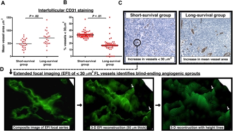
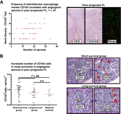
Follicular lymphoma has considerable clinical heterogeneity, and there is a need for easily quantifiable prognostic biomarkers. Microvessel density has been shown to be a useful prognostic factor based on numerical assessment of vessel numbers within histologic sections in some studies, but assessment of tumor neovascularization through angiogenic sprouting may be more relevant. We therefore examined the smallest vessels, single-staining structures measuring less than 30 microm(2) in area, seen within histologic sections, and confirmed that they were neovascular angiogenic sprouts using extended focal imaging. Tissue microarrays composing diagnostic biopsies from patients at the extremes of survival of follicular lymphoma were analyzed with respect to numbers of these sprouts. This analysis revealed higher angiogenic activity in the poor prognostic group and demonstrated an association between increased sprouting and elevated numbers of infiltrating CD163(+) macrophages within the immediate microenvironment surrounding the neovascular sprout.


Figures

Figure 1
Figure 1
Increase in the presence of small vessels in poor prognosis FL identified as angiogenic sprouts using extended focal imaging. (A) CD31 was examined using TMA cores from extremes of survival diagnostic FL biopsies (< 5-year short-survival group and > 15-year long-survival group). Dot-plot chart shows significantly decreased interfollicular CD31+ mean vessel area (μm2) in the short-survival group. Each dot represents an FL patient (mean of triplicate cores) using the TMA. (B) Dot-plot chart shows significantly increased interfollicular CD31+ vascular structures measuring less than 30 μm2 in the short-survival group. Each dot represents a single core (triplicate cores per patient) using the FL extremes of survival TMA. Statistical analysis was performed using a mixed-effects linear model (P values). The black bar represents the mean value for each experimental group. (C) Representative low power (original magnification ×20) images of CD31 interfollicular staining on TMA cores from the short-survival group (left-hand image) and the long-survival group (right-hand image). Note the increase in small, vascular structures measuring less than 30 μm2 in the poor prognosis FL biopsy. The long-survival group exhibited larger mean vessel areas. Image acquisition: Olympus BX61 camera (Applied Imaging), 20×/0.5 numeric aperature (NA); images imported to Ariol Genetix Version 3.2.125 (Applied Imaging). (D) CD31 vascular structures present in the extremes of survival FL TMAs were examined using extended focal imaging (EFI). This technique builds a composite image of a focal series from 50-μm-thick whole sections that can be reconstructed with height lines in 3 dimensions (3-D) using image analysis software (process denoted by black arrows). This EFI analysis identified that the CD31+ vessels measuring less than 30 μm2 in the short-survival FL group (C ●) were blind-ending sprouting processes. The white arrows indicate blind-ending vessels in the poor prognosis FL biopsies identified using EFI. Image acquisition: Olympus BX61 camera (Applied Imaging), 40×/0.75 NA; images constructed by CellF Version 24 (Olympus).
Figure 2
Figure 2
Elevated CD163+ macrophages correlate and are in close proximity with angiogenic sprouts in poor prognosis FL. (A) Dot-plot chart shows a positive correlation (Spearman rank coefficient, r = .43) of interfollicular CD163+ cells (rank density) with the numbers of CD31+ sprouting vessels (< 30 μm2). Representative images are shown on the right-hand side of CD31 and CD163 staining in the short-survival FL group using serial TMA sections. The merge image shows immunofluorescent analysis of these markers using a single section. Image acquisition: Olympus BX61 camera (Applied Imaging), 20×/0.5 NA; images imported to Ariol Genetix Version 3.2.125 (Applied Imaging). (B) Dot-plot chart shows significantly increased CD163+ macrophages in close proximity (within 200 μm) to a CD31+ sprouting vessel in the short-survival FL group compared with the long-survival group and reactive follicular hyperplasia controls. Each dot represents an average number of CD163+ macrophages from 15 analysis areas (200-μm area immediately surrounding a CD31+ sprouting vessel) for each patient sample. The red bar represents the mean value for each experimental group. Statistical analysis was performed using the nonparametric Mann-Whitney test (GraphPad Prism Version 5.03 software; P values). Representative images are shown on the right-hand side of CD31 (labeled yellow using Ariol Genetix Version 3.2.125 analysis software [Applied Imaging]) and CD163 (labeled red) staining in the short- and long-survival FL TMA groups using linked serial TMA sections with Ariol image analysis software. Note the increase numbers of CD163+ macrophages within close proximity (200 μm, black arrow) to a CD31+ sprouting vessel (linked section, blue arrow) in the short-survival group (top images) compared with the long-survival group (bottom images).

References

    1. Johnson PW, Rohatiner AZ, Whelan JS, et al. Patterns of survival in patients with recurrent follicular lymphoma: a 20-year study from a single center. J Clin Oncol. 1995;13(1):140–147. - PubMed
    1. Cullen MH, Lister TA, Brearley RI, Shand WS, Stansfeld AG. Histological transformation of non-Hodgkin's lymphoma: a prospective study. Cancer. 1979;44(2):645–651. - PubMed
    1. Montoto S, Davies AJ, Matthews J, et al. Risk and clinical implications of transformation of follicular lymphoma to diffuse large B-cell lymphoma. J Clin Oncol. 2007;25(17):2426–2433. - PubMed
    1. Solal-Celigny P, Roy P, Colombat P, et al. Follicular lymphoma international prognostic index. Blood. 2004;104(5):1258–1265. - PubMed
    1. Federico M, Bellei M, Marcheselli L, et al. Follicular lymphoma international prognostic index 2: a new prognostic index for follicular lymphoma developed by the international follicular lymphoma prognostic factor project. J Clin Oncol. 2009;27(27):4555–4562. - PubMed

MeSH terms

Substances