Review
. 2010 Apr;23(2):367-81.
doi: 10.1128/CMR.00056-09.

Clinical and laboratory update on blastomycosis

Affiliations
Review

Clinical and laboratory update on blastomycosis

Michael Saccente et al. Clin Microbiol Rev. 2010 Apr.

Abstract

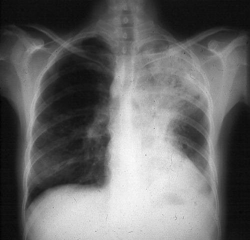
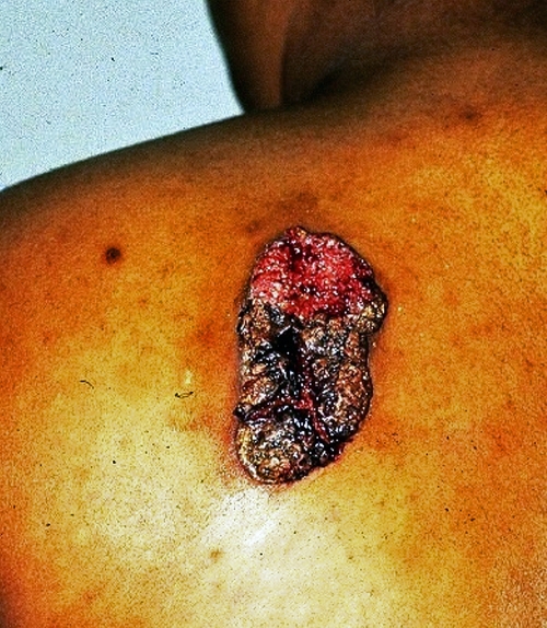
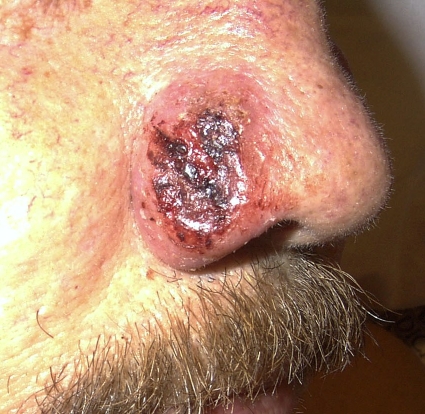
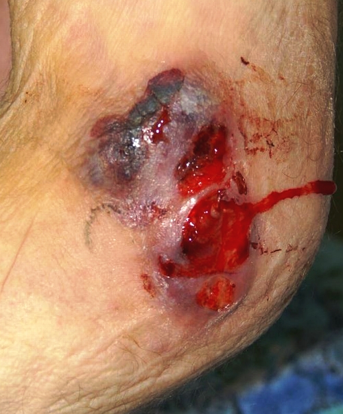
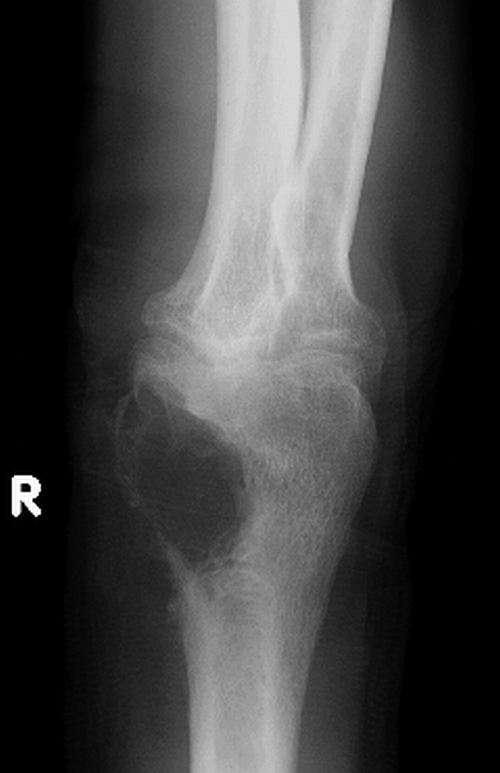
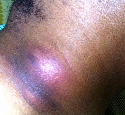
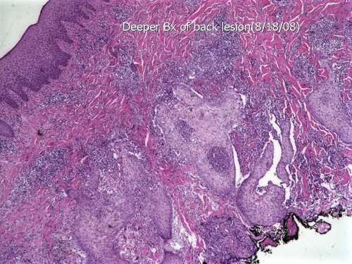
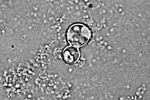
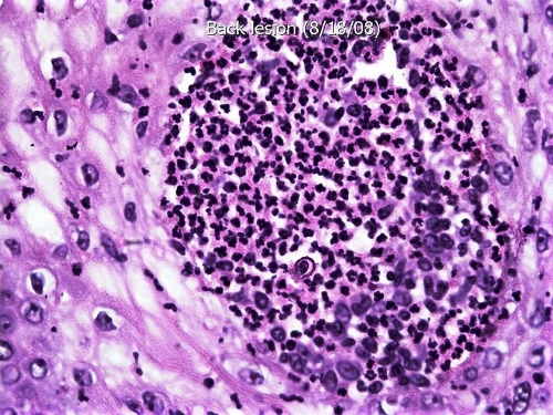
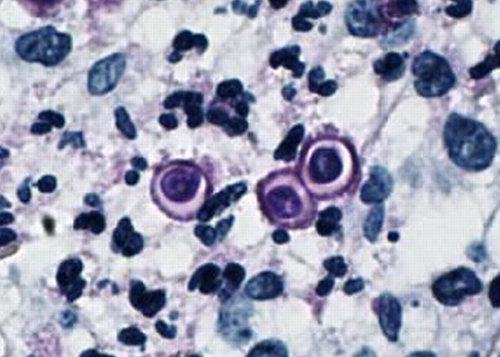
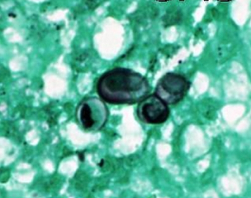
Blastomycosis is endemic in regions of North America that border the Great Lakes and the St. Lawrence River, as well as in the Mississippi River and Ohio River basins. Men are affected more often than women and children because men are more likely to participate in activities that put them at risk for exposure to Blastomyces dermatitidis. Human infection occurs when soil containing microfoci of mycelia is disturbed and airborne conidia are inhaled. If natural defenses in the alveoli fail to contain the infection, lymphohematogenous dissemination ensues. Normal host responses generate a characteristic pyogranulomatous reaction. The most common sites of clinical disease are the lung and skin; osseous, genitourinary, and central nervous system manifestations follow in decreasing order of frequency. Blastomycosis is one of the great mimickers in medicine; verrucous cutaneous blastomycosis resembles malignancy, and mass-like lung opacities due to B. dermatitidis often are confused with cancer. Blastomycosis may be clinically indistinguishable from tuberculosis. Diagnosis is based on culture and direct visualization of round, multinucleated yeast forms that produce daughter cells from a single broad-based bud. Although a long course of amphotericin B is usually curative, itraconazole is also highly effective and is the mainstay of therapy for most patients with blastomycosis.


Figures

FIG. 1.
FIG. 1.
A chest radiograph from a patient with pulmonary blastomycosis showing opacification in the left upper lobe. (Photograph courtesy of Robert W. Bradsher, University of Arkansas for Medical Sciences, Department of Internal Medicine, Division of Infectious Diseases.)
FIG. 2.
FIG. 2.
A verrucous cutaneous lesion due to blastomycosis. Note the irregular shape and crusting. (Photograph courtesy of Robert W. Bradsher, University of Arkansas for Medical Sciences, Department of Internal Medicine, Division of Infectious Diseases.)
FIG. 3.
FIG. 3.
A cutaneous ulcer without exudate on the nose of a renal transplant recipient with blastomycosis. Note the heaped-up borders. (Photograph courtesy of Robert W. Bradsher, University of Arkansas for Medical Sciences, Department of Internal Medicine, Division of Infectious Diseases.)
FIG. 4.
FIG. 4.
An exudative, ulcerative cutaneous lesion on medial aspect of the right elbow of a man with blastomycosis. (Photograph courtesy of J. Ryan Bariola, University of Arkansas for Medical Sciences, Department of Internal Medicine, Division of Infectious Diseases.)
FIG. 5.
FIG. 5.
Radiograph of the right arm shown in Fig. 4 that reveals a large osteolytic lesion of the distal humerus due to osteomyelitis. R, right. (Photograph courtesy of J. Ryan Bariola, University of Arkansas for Medical Sciences, Department of Internal Medicine, Division of Infectious Diseases.)
FIG. 6.
FIG. 6.
A subcutaneous neck mass with overlying cutaneous erythema due to blastomycosis. (Photograph courtesy of Robert W. Bradsher, University of Arkansas for Medical Sciences, Department of Internal Medicine, Division of Infectious Diseases.)
FIG. 7.
FIG. 7.
Hematoxylin-and-eosin-stained section of an ulcerated lesion on the thigh shows pseudoepitheliomatous hyperplasia, epidermal abscesses, and a prominent inflammatory infiltrate in the dermis (low-power magnification). Bx, biopsy. (Photograph courtesy of Jameel Ahmad Brown, University of Arkansas for Medical Sciences, Department of Pathology.)
FIG. 8.
FIG. 8.
KOH preparation of clinical material showing the typical appearance of B. dermatitidis. Note the round shape, doubly refractile wall, and single broad-based bud. (Photograph courtesy of Robert W. Bradsher, University of Arkansas for Medical Sciences, Department of Internal Medicine, Division of Infectious Diseases.)
FIG. 9.
FIG. 9.
High-power magnification of the section illustrated in Fig. 7 shows a microabscess with a nonbudding yeast cell of B. dermatitidis. Note the characteristic thick cell wall and centrally retracted cytoplasm (high-power magnification). (Photograph courtesy of Jameel Ahmad Brown, University of Arkansas for Medical Sciences, Department of Pathology.)
FIG. 10.
FIG. 10.
PAS-stained section of the lesion illustrated in Fig. 7 shows a budding yeast of B. dermatitidis with the characteristic broad-based bud, as well as a single nonbudding yeast cell (high-power magnification). (Photograph courtesy of Jameel Ahmad Brown, University of Arkansas for Medical Sciences, Department of Pathology.)
FIG. 11.
FIG. 11.
GMS-stained section of the lesion illustrated in Fig. 7 shows a budding yeast of B. dermatitidis with the characteristic broad-based bud, as well as two slightly misshapen single nonbudding yeast cells (high-power magnification). (Photograph courtesy of Jameel Ahmad Brown, University of Arkansas for Medical Sciences, Department of Pathology.)

References

    1. Abernathy, R. S. 1959. Clinical manifestations of pulmonary blastomycosis. Ann. Intern. Med. 51:707-727. - PubMed
    1. Abernathy, R. S. 1967. Amphotericin therapy of North American blastomycosis. Antimicrob. Agents Chemother. 3:208-211. - PubMed
    1. Amini, M., W. E. Shams, T. Barklow, and F. A. Sarubbi. 2007. A 45-year-old man with fever and adult respiratory distress syndrome. Clin. Infect. Dis. 44:566-567, 615-616. - PubMed
    1. Ashley, E. S. D., R. Lewis, J. S. Lewis, C. Martin, and D. Andes. 2006. Pharmacology of systemic antifungal agents. Clin. Infect. Dis. 43:S28-S39.
    1. Axelson, G. K., T. Giorgadze, and G. A. Youngberg. 2008. Evaluation of the use of Congo red staining in the differential diagnosis of Candida vs. various other yeast-form fungal organisms. J. Cutan. Pathol. 35:27-30. - PubMed

LinkOut - more resources