. 2009;1(1):29-45.
doi: 10.1159/000142731. Epub 2008 Jul 2.

Attenuated activation of macrophage TLR9 by DNA from virulent mycobacteria

Affiliations

Attenuated activation of macrophage TLR9 by DNA from virulent mycobacteria

Alexandra K Kiemer et al. J Innate Immun. 2009.

Abstract

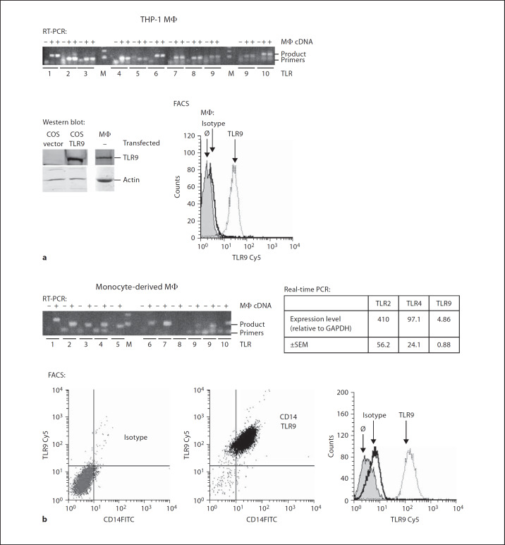
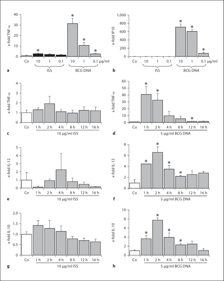
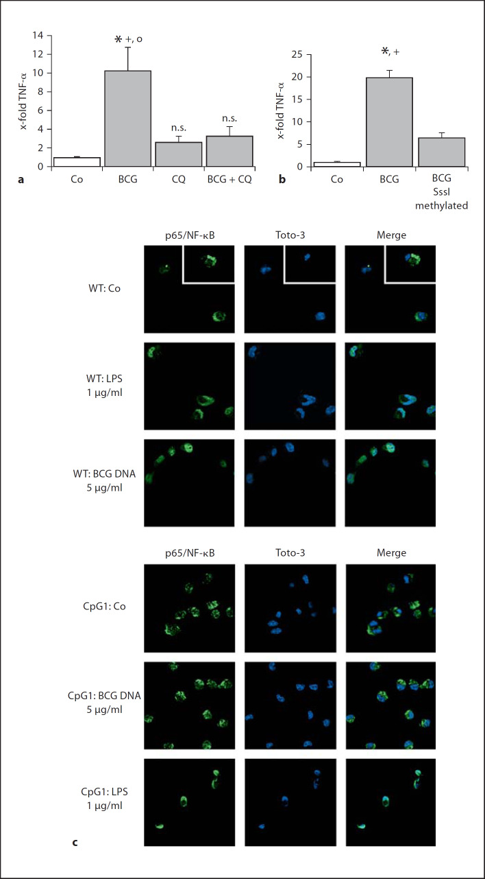
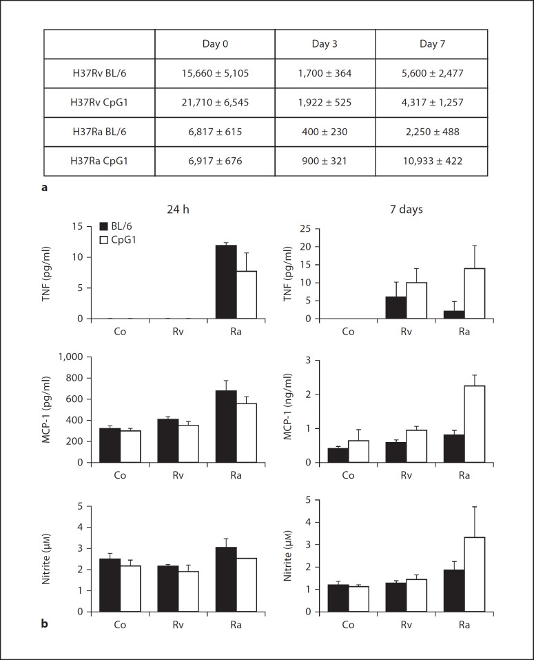
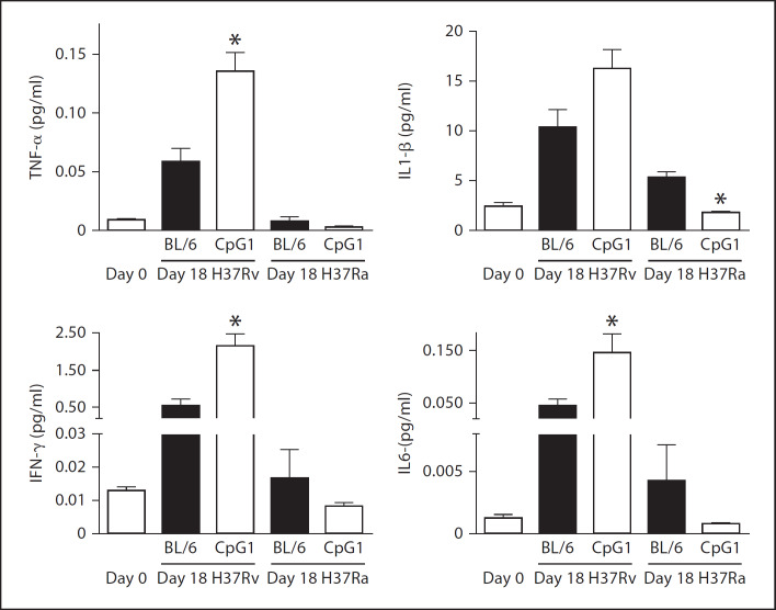
Alveolar macrophages are the first line of host defence against mycobacteria, but an insufficient host response allows survival of bacteria within macrophages. We aimed to investigate the role of Toll-like receptor 9 (TLR9) activation in macrophage defence against mycobacteria. Human in vitro differentiated macrophages as well as human and mouse alveolar macrophages showed TLR9 mRNA and protein expression. The cells were markedly activated by DNA isolated from attenuated mycobacterial strains (H37Ra and Mycobacterium bovis BCG) as assessed by measuring cytokine expression by real-time PCR, whereas synthetic phosphorothioate-modified oligonucleotides had a much lower potency to activate human macrophages. Intracellular replication of H37Ra was higher in macrophages isolated from TLR9-deficient mice than in macrophages from wild-type mice, whereas H37Rv showed equal survival in cells from wild-type or mutant mice. Increased bacterial survival in mouse macrophages was accompanied by altered cytokine production as determined by Luminex bead assays. In vivo infection experiments also showed differential cytokine production in TLR9-deficient mice compared to wild-type animals. Both human monocyte-derived macrophages as well as human alveolar macrophages showed reduced activation upon treatment with DNA isolated from bacteria from virulent (M. bovis and H37Rv) compared to attenuated mycobacteria. We suggest attenuated TLR9 activation contributes to the insufficient host response against virulent mycobacteria.


Figures

Fig. 1
Fig. 1
Human monocyte-derived macrophages express TLR9. THP-1 cells were differentiated by PMA (a) and primary monocytes were differentiated into macrophages by M-CSF treatment (b). RT-PCR and real-time PCR: RNA isolation, reverse transcription and qualitative RT-PCR for TLRs1–10 as well as quantitative real-time PCR for TLR2, TLR4 and TLR9 were performed as described in Materials and Methods. Qualitative RT-PCR was performed in the presence (+) or absence (–) of cDNA. Reaction products of qualitative RT-PCR reactions and a size marker (M) were electrophoresed in agarose and stained with ethidium bromide. Expression levels of TLR2, TLR4 and TLR9 are expressed as mRNA copies of the respective gene per 1,000 copies of GAPDH. Quantification was performed by comparing starting amounts of cDNAs with a standard curve of amplicon cloned into pGEM-T Easy. Data represent means ± SEM of cells from 5–7 donors. Western blot: COS-1 cells were transfected with a pcDNA3.1 vector encoding TLR9 or a vector control. Transfected cells as well as human macrophages were lysed. TLR9 was detected by Western blot by a custom antibody and visualized by an IRDye 800 conjugated anti-mouse IgG. Actin served as a loading control. FACS: Primary macrophages and differentiated THP-1 cells were stained with a custom antibody against TLR9 followed by detection with a secondary antibody via biotin/streptavidin/Cy5 and an FITC-labelled commercial antibody against CD14 as described in Materials and Methods. Cells were analysed by flow cytometry. Data show 1 representative of 3 independent experiments with cells from different donors.
Fig. 2
Fig. 2
Activation of human monocyte-derived macrophages by mycobacterial DNA. Primary monocyte-derived macrophages were left untreated (Co) or treated with ISS (ISS 1018, phosphorothioate-modified oligonucleotide 5’-TGACTGTGAACGTTCGAGATGA-3’) or genomic DNA from M. bovis BCG. a, b Cells were treated for 3 h in the indicated concentrations (0.1–10 µg/ml). c, d Cells were treated for the indicated times (1–16 h) in the indicated concentrations (5 or 10 µg/ml). After treatment, RNA was isolated followed by real-time PCR analysis for TNF-α (a, c, d), IP10 (b), IL-12 (e, f) or IL-10 (g, h). Values for cytokines were normalized to the housekeeping gene GAPDH. Data are expressed as x-fold TNF-α or IP10 induction with expression levels of untreated cells set as 1. Data show means ± SEM of 3 experiments performed in triplicates. * p < 0.05 significantly different from Co (t test).
Fig. 3
Fig. 3
Activation by DNA preparations is specific for non-methylated DNA. a, b DNA was isolated from M. bovis BCG mycobacteria and added to primary monocyte-derived macrophages for 3 h followed by RNA isolation and real-time PCR analysis for TNF-α. a Cells were either left untreated (Co), or treated with CQ (10 µM), BCG DNA (5 µg/ml) or a combination of both, whereby CQ was added to the cells 1 h before BCG DNA. * p < 0.05 significantly different from Co; + p < 0.05 significantly different from CQ; ° p < 0.05 significantly different from BCG + CQ. n.s. = Not significantly different from Co (t test). b DNA from BCG mycobacteria was methylated with SssI methylase. In vitro differentiated macrophages were either left untreated (Co) or treated with methylated or mock-treated DNA. Expression levels for TNF-α were normalized on housekeeping genes and data are expressed as x-fold TNF-α induction. Data show means ± SEM of 3 experiments, each performed in triplicates. * p < 0.05 significantly different from Co; + p < 0.05 significantly different from BCG SssI methylated (t test). c Mouse alveolar macrophages from wild-type (WT) or TLR9CpG1/CpG1 (CpG1) mice were either left untreated (Co) or treated with LPS (1 µg/ml) or BCG DNA (5 µg/ml) before staining of p65/NF-κB (green) and nuclear staining with Toto-3 (blue) as described in Materials and Methods. Data show channels for each of the single fluorophores (left and middle panel) and a merged overlay (right panel).
Fig. 4
Fig. 4
Human alveolar macrophages express TLR9 mRNA and protein. TLR mRNA expression was analysed in human alveolar macrophages by qualitative (a) and quantitative RT-PCR (b) and TLR9 protein was detected by immunocytochemistry and Western blot (c). a, b RNA isolation, reverse transcription and qualitative RT-PCR for TLR2 and TLR9 as well as quantitative PCR for TLR2, TLR4 and TLR9 were performed as described in Materials and Methods. Qualitative RT-PCR was performed in the presence (+) or absence (–) of cDNA. Reaction products of qualitative RT-PCR reactions and a size marker (M) were electrophoresed and stained with ethidium bromide. Expression levels of TLR2, TLR4 and TLR9 are expressed as mRNA copies of the respective gene per 1,000 copies of GAPDH. Quantification was performed by comparing starting amounts of cDNAs with a standard curve of amplicon cloned into pGEM-T Easy. c Alveolar macrophages were stained for TLR9 (blue) and with propidium iodide (red) as described in Materials and Methods employing a custom-made antibody and a secondary antibody detected via biotin/streptavidin-Cy5. Stained cells were analysed by confocal microscopy and laser scanning cytometry. A laser scanning cytometer equipped with an argon-ion air-cooled laser and an HeNe laser equipped with a digital camera were used to measure Cy5 fluorescence. Nuclear propidium iodide fluorescence was used as the contouring parameter. Alveolar macrophages for the different analyses were each obtained from different donors. For Western blots, PMA-differentiated THP-1 macrophages or alveolar macrophages were lysed. TLR9 was detected by Western blot by a custom antibody and visualized by an IRDye 800 conjugated anti-mouse IgG. Actin served as a loading control.
Fig. 5
Fig. 5
Mycobacterial infection of murine macrophages. Mouse bone marrow macrophages were derived from wild-type (BL/6) or TLR9CpG1/CpG1 mice (CpG1) and were either mock infected (Co) or infected with virulent H37Rv or attenuated H37Ra. a Cells were lysed 3 and 7 days after infection and plated in serial dilutions on 7H11 agar to enumerate CFU. Data show values from 2 independent experiments with 3–6 infections (means ± SEM, cell preparations from 6–9 mice per strain). b Cell supernatants from mock-infected cells (Co) or from cells infected with virulent H37Rv or attenuated H37Ra were taken off at days 1 and 7 after infection and TNF-α, MCP-1 and nitrite levels were determined as described in Materials and Methods. Data are expressed as means ± SD of cell preparations from 6–9 mice per strain, performed in triplicates.
Fig. 6
Fig. 6
Altered cytokine production in TLR9-deficient animals infected with virulent or attenuated mycobacteria. Wild-type (BL/6) or TLR9CpG1/CpG1 mice (CpG1) were infected with M. tuberculosis H37Rv or H37Ra. Lungs were homogenized on day 0 or day 18 and TNF-α, IL-1β, IFN-γ and IL-6 levels were measured by a Luminex bead assay (Upstate Beadlyte). Data show means ± SEM of lungs from 4 animals. * p < 0.05 statistically different from values in wild-type animals.
Fig. 7
Fig. 7
Activation of human monocyte-derived and alveolar macrophages by DNA from virulent and attenuated mycobacteria. Monocyte-derived (a) or alveolar (b) macrophages were treated by DNA from virulent M. tuberculosis (H37Rv) and M. bovis or from attenuated H37Ra and M. bovis BCG strains for 3 h (a) or 2 h (b). RNA was isolated, followed by real-time PCR analysis for TNF-α. Data are expressed as TNF-α induction after normalization to housekeeping gene expression. Data show means ± SEM of 3–13 values employing cells from 3–5 different donors. * p < 0.05 significant difference between treatment with the same DNA concentration of a virulent and the respective attenuated strain (t test). c DNA was isolated from H37Ra mycobacteria and digested with DNase. Primary monocyte-derived macrophages were either left untreated (Co) or treated with digested or mock-treated DNA followed by RNA isolation and real-time PCR analysis for TNF-α. Data are expressed as x-fold TNF-α induction after normalization to GAPDH values. Data show means ± SEM of 3 independent experiments performed in triplicate. * p < 0.05 significantly different to Co; + p < 0.05 significantly different to DNase digested (t test).

Similar articles

Cited by

References

    1. Doffinger R, Patel SY, Kumararatne DS. Host genetic factors and mycobacterial infections: lessons from single gene disorders affecting innate and adaptive immunity. Microbes Infect. 2006;8:1141–1150. - PubMed
    1. Beutler B. Inferences, questions and possibilities in Toll-like receptor signalling. Nature. 2004;430:257–263. - PubMed
    1. Shimada S, Yano O, Inoue H, Kuramoto E, Fukuda T, Yamamoto H, Kataoka T, Tokunaga T. Antitumor activity of the DNA fraction from Mycobacterium bovis BCG. II. Effects on various syngeneic mouse tumors. J Natl Cancer Inst. 1985;74:681–688. - PubMed
    1. Tokunaga T, Yamamoto H, Shimada S, Abe H, Fukuda T, Fujisawa Y, Furutani Y, Yano O, Kataoka T, Sudo T. Antitumor activity of deoxyribonucleic acid fraction from Mycobacterium bovis BCG. I. Isolation, physicochemical characterization, and antitumor activity. J Natl Cancer Inst. 1984;72:955–962. - PubMed
    1. Bafica A, Scanga CA, Feng CG, Sher A, Leifer C, Cheever A. TLR9 regulates Th1 responses and cooperates with TLR2 in mediating optimal resistance to Mycobacterium tuberculosis. J Exp Med. 2005;202:1715–1724. - PMC - PubMed

Publication types

MeSH terms