

Survival of bacterial biofilms within neutrophil extracellular traps promotes nontypeable Haemophilus influenzae persistence in the chinchilla model for otitis media
- PMID: 20375579
- PMCID: PMC6951045
- DOI: 10.1159/000205937
Survival of bacterial biofilms within neutrophil extracellular traps promotes nontypeable Haemophilus influenzae persistence in the chinchilla model for otitis media
Abstract
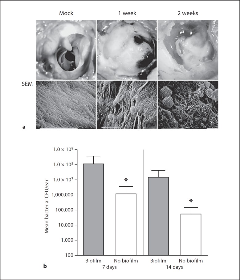
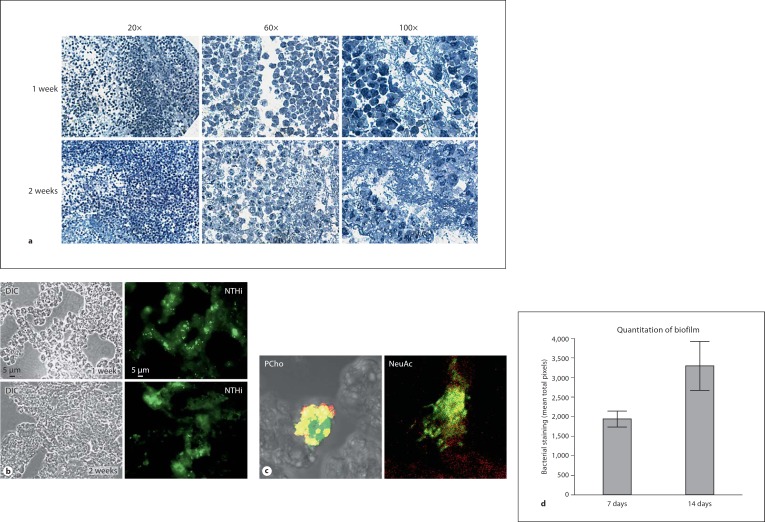
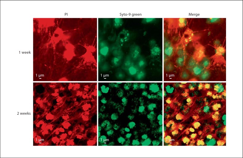
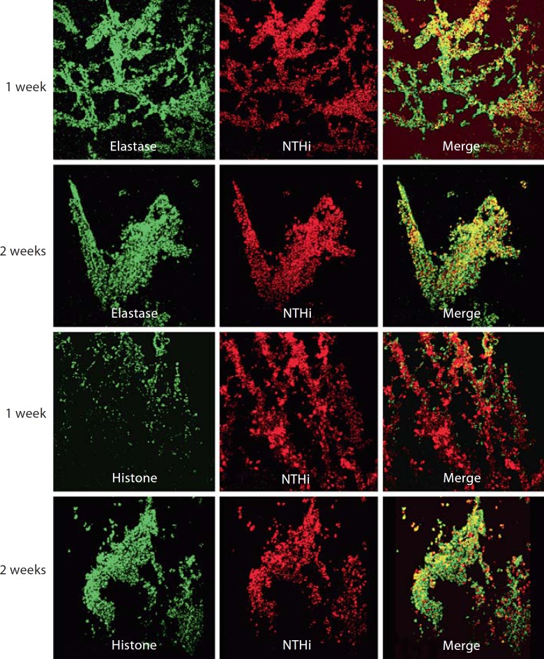
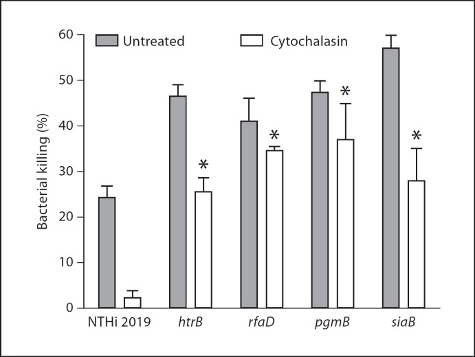
Nontypeable Haemophilus influenzae (NTHi) is a leading cause of acute and chronic otitis media, which are a major public health problem worldwide. The persistence of NTHi during chronic and recurrent otitis media infections involves multicellular biofilm communities formed within the middle-ear chamber. Bacterial biofilms resist immune clearance and antibiotic therapy due in part to encasement within a polymeric matrix. In this study, the contribution of biofilms to bacterial persistence in vivo and composition of the NTHi biofilm matrix during experimental otitis media were investigated. The presence of biofilms within the chinchilla middle-ear chamber was significantly correlated with increased bacterial load in middle-ear effusions and tissue. Examination of thin sections revealed polymorphonuclear cells within a DNA lattice containing elastase and histones, which is consistent with the definition of neutrophil extracellular traps. Viable multicellular biofilm communities with biofilm phenotypes were found within the DNA lattice throughout the biofilm. Further, NTHi was resistant to both phagocytic and extracellular neutrophil killing in vitro by means of lipooligosaccharide moieties that promote biofilm formation. These data support the conclusion that NTHi subverts neutrophil extracellular traps to persist in vivo. These data also indicate that a more inclusive definition for biofilms may be warranted.
Copyright 2009 S. Karger AG, Basel.
Figures
Similar articles
-
Phosphorylcholine decreases early inflammation and promotes the establishment of stable biofilm communities of nontypeable Haemophilus influenzae strain 86-028NP in a chinchilla model of otitis media.Infect Immun. 2007 Feb;75(2):958-65. doi: 10.1128/IAI.01691-06. Epub 2006 Nov 27. Infect Immun. 2007. PMID: 17130253 Free PMC article.
-
Phosphorylcholine expression by nontypeable Haemophilus influenzae correlates with maturation of biofilm communities in vitro and in vivo.J Bacteriol. 2007 Nov;189(22):8300-7. doi: 10.1128/JB.00532-07. Epub 2007 Jun 15. J Bacteriol. 2007. PMID: 17573475 Free PMC article.
-
Dps promotes survival of nontypeable Haemophilus influenzae in biofilm communities in vitro and resistance to clearance in vivo.Front Cell Infect Microbiol. 2012 May 3;2:58. doi: 10.3389/fcimb.2012.00058. eCollection 2012. Front Cell Infect Microbiol. 2012. PMID: 22919649 Free PMC article.
-
Novel concepts in nontypeable Haemophilus influenzae biofilm formation.FEMS Microbiol Lett. 2013 Sep;346(2):81-9. doi: 10.1111/1574-6968.12203. Epub 2013 Jul 15. FEMS Microbiol Lett. 2013. PMID: 23808954 Review.
-
Nontypeable Haemophilus influenzae biofilms: role in chronic airway infections.Front Cell Infect Microbiol. 2012 Jul 25;2:97. doi: 10.3389/fcimb.2012.00097. eCollection 2012. Front Cell Infect Microbiol. 2012. PMID: 22919686 Free PMC article. Review.
Cited by
-
The Haemophilus influenzae Sap transporter mediates bacterium-epithelial cell homeostasis.Infect Immun. 2013 Jan;81(1):43-54. doi: 10.1128/IAI.00942-12. Epub 2012 Oct 15. Infect Immun. 2013. PMID: 23071138 Free PMC article.
-
Mimicking biofilm formation and development: Recent progress in in vitro and in vivo biofilm models.iScience. 2021 Apr 17;24(5):102443. doi: 10.1016/j.isci.2021.102443. eCollection 2021 May 21. iScience. 2021. PMID: 34013169 Free PMC article. Review.
-
Optimizing synthetic cystic fibrosis sputum media for growth of non-typeable Haemophilus influenzae.Access Microbiol. 2025 Jun 20;7(6):000979.v3. doi: 10.1099/acmi.0.000979.v3. eCollection 2025. Access Microbiol. 2025. PMID: 40548129 Free PMC article.
-
Lipopolysaccharide Biosynthesis Genes of Yersinia pseudotuberculosis Promote Resistance to Antimicrobial Chemokines.PLoS One. 2016 Jun 8;11(6):e0157092. doi: 10.1371/journal.pone.0157092. eCollection 2016. PLoS One. 2016. PMID: 27275606 Free PMC article.
-
Autoinducer 2 (AI-2) Production by Nontypeable Haemophilus influenzae 86-028NP Promotes Expression of a Predicted Glycosyltransferase That Is a Determinant of Biofilm Maturation, Prevention of Dispersal, and Persistence In Vivo.Infect Immun. 2018 Nov 20;86(12):e00506-18. doi: 10.1128/IAI.00506-18. Print 2018 Dec. Infect Immun. 2018. PMID: 30249749 Free PMC article.
References
-
- Klein JO. The burden of otitis media. Vaccine. 2000;19:S2–S8. - PubMed
-
- Bakaletz LO. Bacterial biofilms in otitis media: evidence and relevance. Pediatr Infect Dis J. 2007;26:S17–S19. - PubMed
-
- Fux CA, Costerton JW, Stewart PS, Stoodley P. Survival strategies of infectious biofilms. Trends Microbiol. 2005;13:34–40. - PubMed
Publication types
MeSH terms
Substances
Grants and funding
LinkOut - more resources
Full Text Sources
Medical
