

Evidence for altered Wnt signaling in psoriatic skin
- PMID: 20376066
- PMCID: PMC2886156
- DOI: 10.1038/jid.2010.67
Evidence for altered Wnt signaling in psoriatic skin
Abstract
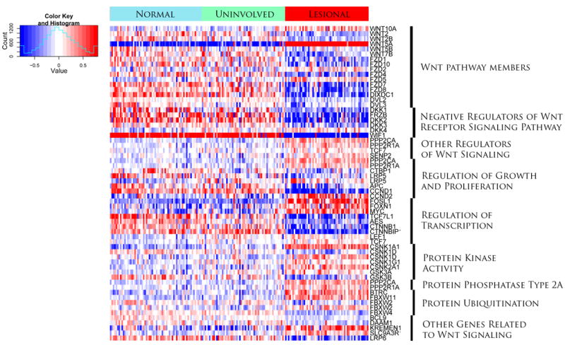
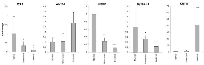
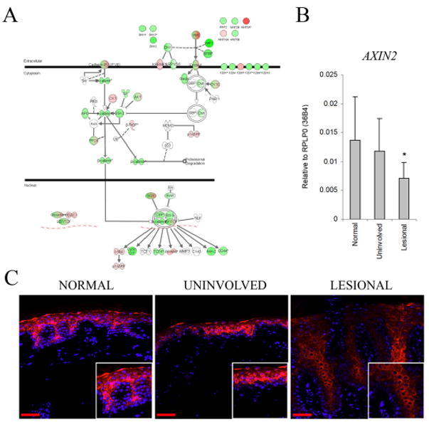
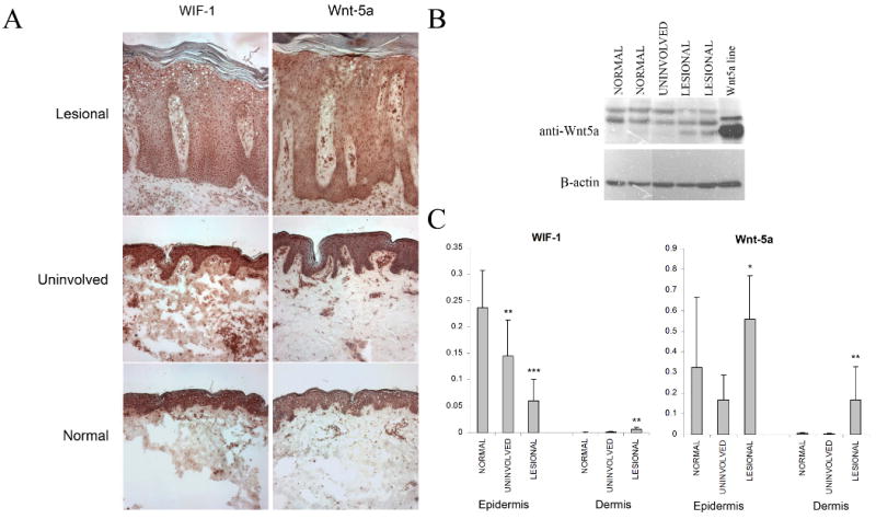
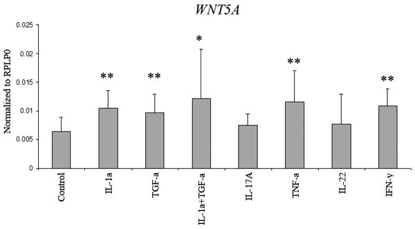
The Wnt gene family encodes a set of highly conserved secreted signaling proteins that have major roles in embryogenesis and tissue homeostasis. Yet the expression of this family of important mediators in psoriasis, a disease characterized by marked changes in keratinocyte growth and differentiation, is incompletely understood. We subjected 58 paired biopsies from lesional and uninvolved psoriatic skin and 64 biopsies from normal skin to global gene expression profiling. WNT5A transcripts were upregulated fivefold in lesional skin, accompanied by increased Wnt-5a protein levels. Notably, WNT5A mRNA was markedly induced by IL-1alpha, tumor necrosis factor-alpha, IFN-gamma, and transforming growth factor-alpha in cultured keratinocytes. Frizzled 2 (FZD2) and FZD5, which encode receptors for Wnt5A, were also increased in lesional psoriatic skin. In contrast, expression of WIF1 mRNA, encoding a secreted antagonist of the Wnt proteins, was downregulated >10-fold in lesional skin, along with decreased WNT inhibitory factor (WIF)-1 immunostaining. Interestingly, pathway analysis along with reduced AXIN2 expression and lack of nuclear translocation of beta-catenin indicated a suppression of canonical Wnt signaling in lesional skin. The results of our study suggest a shift away from canonical Wnt signaling toward noncanonical pathways driven by interactions between Wnt-5a and its cognate receptors in psoriasis, accompanied by impaired homeostatic inhibition of Wnt signaling by WIF-1 and dickkopf.
Figures
Similar articles
-
Wnt/β-Catenin and Wnt5a/Ca Pathways Regulate Proliferation and Apoptosis of Keratinocytes in Psoriasis Lesions.Cell Physiol Biochem. 2015;36(5):1890-902. doi: 10.1159/000430158. Epub 2015 Jul 17. Cell Physiol Biochem. 2015. PMID: 26202350
-
Transcriptional profiling shows altered expression of wnt pathway- and lipid metabolism-related genes as well as melanogenesis-related genes in melasma.J Invest Dermatol. 2011 Aug;131(8):1692-700. doi: 10.1038/jid.2011.109. Epub 2011 May 12. J Invest Dermatol. 2011. PMID: 21562572
-
Increased expression of Wnt5a in psoriatic plaques.J Invest Dermatol. 2007 Jan;127(1):163-9. doi: 10.1038/sj.jid.5700488. Epub 2006 Jul 20. J Invest Dermatol. 2007. PMID: 16858420
-
Hear the Wnt Ror: how melanoma cells adjust to changes in Wnt.Pigment Cell Melanoma Res. 2009 Dec;22(6):724-39. doi: 10.1111/j.1755-148X.2009.00627.x. Epub 2009 Aug 25. Pigment Cell Melanoma Res. 2009. PMID: 19708915 Free PMC article. Review.
-
Secreted antagonists of the Wnt signalling pathway.J Cell Sci. 2003 Jul 1;116(Pt 13):2627-34. doi: 10.1242/jcs.00623. J Cell Sci. 2003. PMID: 12775774 Review.
Cited by
-
Assessment of ADAM17 and ADAM10 proteins with CXCL10 and thyroid autoimmunity in vitiligo pathogenesis.Postepy Dermatol Alergol. 2022 Apr;39(2):397-400. doi: 10.5114/ada.2022.115891. Epub 2022 May 9. Postepy Dermatol Alergol. 2022. PMID: 35645671 Free PMC article.
-
Logical and experimental modeling of cytokine and eicosanoid signaling in psoriatic keratinocytes.iScience. 2021 Nov 15;24(12):103451. doi: 10.1016/j.isci.2021.103451. eCollection 2021 Dec 17. iScience. 2021. PMID: 34877506 Free PMC article.
-
Activation of Transcription Factor 4 in Dendritic Cells Controls Th1/Th17 Responses and Autoimmune Neuroinflammation.J Immunol. 2021 Sep 1;207(5):1428-1436. doi: 10.4049/jimmunol.2100010. Epub 2021 Aug 4. J Immunol. 2021. PMID: 34348977 Free PMC article.
-
Canonical wnt signaling in dendritic cells regulates Th1/Th17 responses and suppresses autoimmune neuroinflammation.J Immunol. 2015 Apr 1;194(7):3295-304. doi: 10.4049/jimmunol.1402691. Epub 2015 Feb 20. J Immunol. 2015. PMID: 25710911 Free PMC article.
-
Transcriptome Analysis of Host Inflammatory Responses to the Ectoparasitic Mite Sarcoptes scabiei var. hominis.Front Immunol. 2021 Dec 1;12:778840. doi: 10.3389/fimmu.2021.778840. eCollection 2021. Front Immunol. 2021. PMID: 34925353 Free PMC article.
References
-
- Ahumada A, Slusarski DC, Liu X, Moon RT, Malbon CC, Wang HY. Signaling of rat Frizzled-2 through phosphodiesterase and cyclic GMP. Science. 2002;298:2006–10. - PubMed
-
- Belso N, Szell M, Pivarcsi A, Kis K, Kormos B, Kenderessy AS, et al. Differential expression of D-type cyclins in HaCaT keratinocytes and in psoriasis. J Invest Dermatol. 2008;128:634–42. - PubMed
-
- Blumenthal A, Ehlers S, Lauber J, Buer J, Lange C, Goldmann T, et al. The Wingless homolog WNT5A and its receptor Frizzled-5 regulate inflammatory responses of human mononuclear cells induced by microbial stimulation. Blood. 2006;108:965–73. - PubMed
-
- Cheng CW, Yeh JC, Fan TP, Smith SK, Charnock-Jones DS. Wnt5a-mediated non-canonical Wnt signalling regulates human endothelial cell proliferation and migration. Biochem Biophys Res Commun. 2008;365:285–90. - PubMed
Publication types
MeSH terms
Substances
Grants and funding
LinkOut - more resources
Full Text Sources
Other Literature Sources
Medical
