Comparison of proliferative and multilineage differentiation potentials of cord matrix, cord blood, and bone marrow mesenchymal stem cells
- PMID: 20376261
- PMCID: PMC2847339
- DOI: 10.4103/0973-6247.59386
Comparison of proliferative and multilineage differentiation potentials of cord matrix, cord blood, and bone marrow mesenchymal stem cells
Abstract
Background: Hematopoietic stem cells (HSCs) and mesenchymal stem cells (MSCs) are the two widely studied and characterized adult stem cells. Thus far, MSCs were obtained from the bone marrow, which is a painful procedure. Therefore, MSCs from less common sources like cord blood, adipose tissue, tooth pulp, and so on, have been the subject of research. The purpose of this study is to explore the possibility of finding MSCs from a less controversial, easy, and abundant source, such as the umbilical cord, for potential regenerative medicine applications.
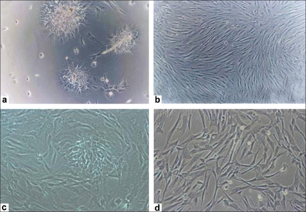
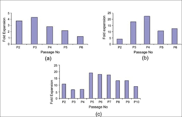
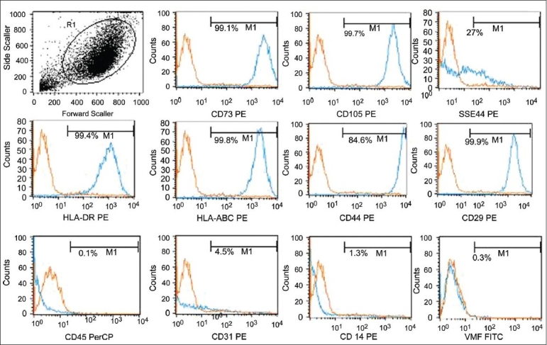
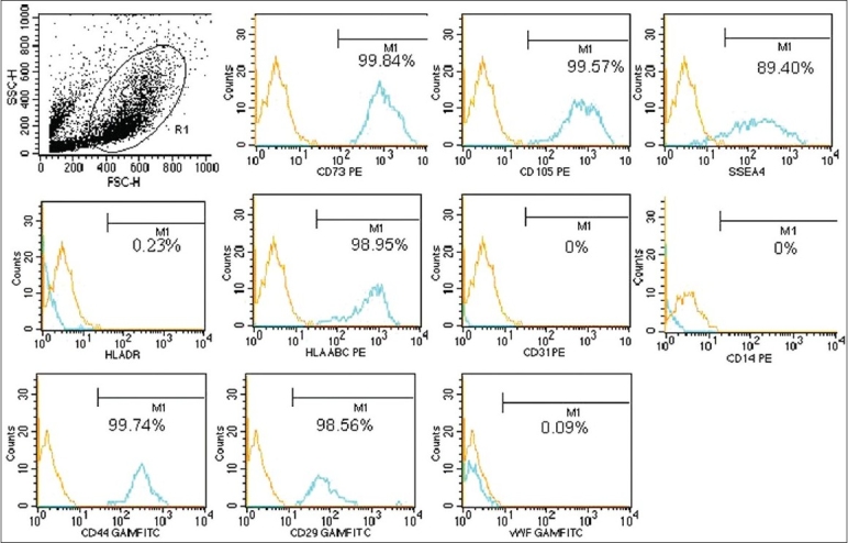
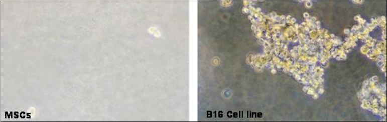
Study design and methods: Five bone marrow samples (BM), seventy cord blood units (CB), and four umbilical cord matrix (CM) samples have been used for the study. Expanded MSCs were checked for biomarker expression by flow cytometry and were also checked for their differentiation to mesodermal and ectodermal lineages.
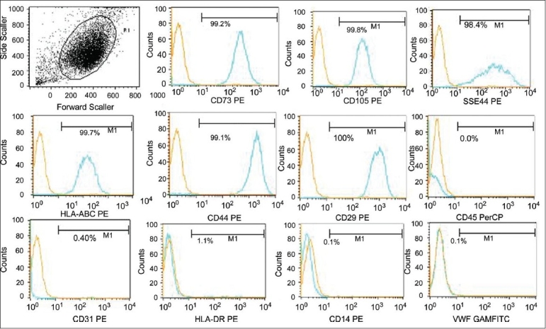
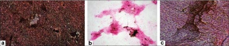
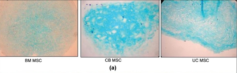
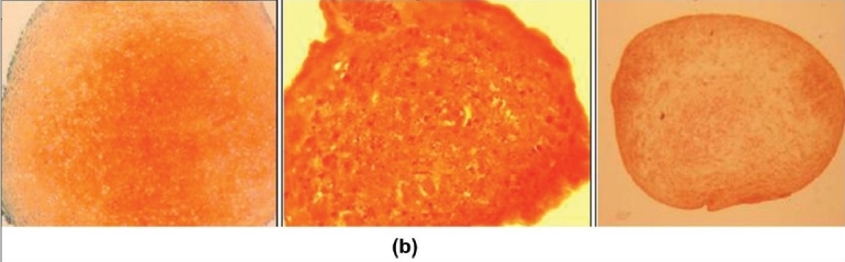
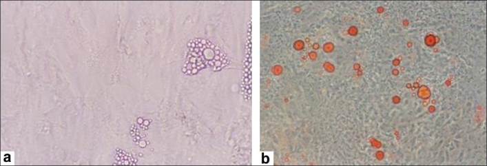
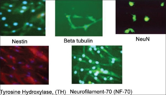
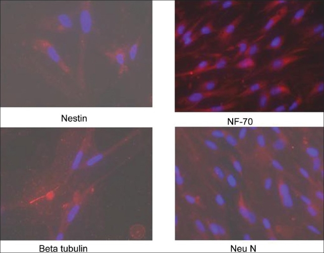
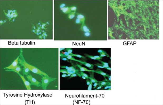
Results: MSCs could be isolated from 100% BM and CM samples, as compared to only 6% of CB samples. The fold expansion of the mesenchymal stem cells observed in CB (CB-MSCs) was distinctly higher as compared to BM (BM-MSCs) and CM (CM-MSCs). MSCs isolated from all the three sources expressed a characteristic mesenchymal phenotype of CD45 - /vWF - /CD14 - /CD31 - /CD73 + /CD105 + /SSEA4 + /CD29 + /CD44 + /HLAABC +, whereas, the HLA DR was conspicuously absent in CM-MSCs and CB-MSCs. Although osteogenic, chondrogenic, and neural differentiation was observed in MSCs from all sources, adipogenic differentiation was observed only in BM-MSCs.
Conclusion: CM-MSCs are a dependable source of an unlimited number of MSCs for autologous and allogenic use in regenerative medicine.
Keywords: Mesenchymal stem cells; bone marrow; umbilical cord; umbilical cord blood.
Conflict of interest statement
Figures
References
-
- Adel A, Jeremy Mao J. Mesenchymal Stem Cells: Isolation and Therapeutics. Stem Cells and Devel. 2004;13:436–48. - PubMed
-
- Pittenger MF, Mackay AM, Beck SC, Jaiswal RK, Douglas R, Mosca JD, et al. Multilineage potential of adult human mesenchymal stem cells. Science. 1999;284:143–7. - PubMed
-
- Frank PB, J Mary M. Mesenchymal stem cells: Clinical applications and biological characterization. Int J of Biochem and Cell Bio. 2004:568–84. - PubMed
-
- D'Ippolito G, Schiller PC, Ricordi C, Roos BA, Howard GA. Age-related osteogenic potential of mesenchymal stromal stem cells from human vertebral bone marrow. J Bone Miner Res. 1999;14:1115–22. - PubMed
-
- Kern S, Eichler H, Stoeve J, Klüter H, Bieback K. Comparative analysis of mesenchymal stem cells from bone marrow, umbilical cord blood, or adipose tissue. Stem Cells. 2006;24:1294–301. - PubMed
LinkOut - more resources
Full Text Sources
Other Literature Sources
Research Materials
Miscellaneous