. 2010 Apr 1;5(4):e9981.
doi: 10.1371/journal.pone.0009981.

The combinatorial PP1-binding consensus Motif (R/K)x( (0,1))V/IxFxx(R/K)x(R/K) is a new apoptotic signature

Affiliations

The combinatorial PP1-binding consensus Motif (R/K)x( (0,1))V/IxFxx(R/K)x(R/K) is a new apoptotic signature

Angélique N Godet et al. PLoS One. .

Abstract

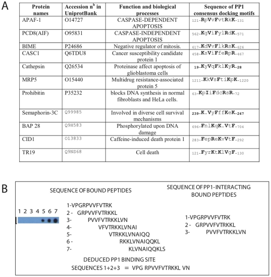
Background: Previous studies established that PP1 is a target for Bcl-2 proteins and an important regulator of apoptosis. The two distinct functional PP1 consensus docking motifs, R/Kx((0,1))V/IxF and FxxR/KxR/K, involved in PP1 binding and cell death were previously characterized in the BH1 and BH3 domains of some Bcl-2 proteins.

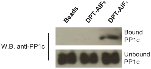
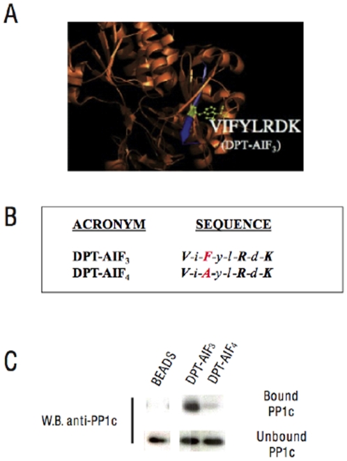
Principal findings: In this study, we demonstrate that DPT-AIF(1), a peptide containing the AIF(562-571) sequence located in a c-terminal domain of AIF, is a new PP1 interacting and cell penetrating molecule. We also showed that DPT-AIF(1) provoked apoptosis in several human cell lines. Furthermore, DPT-APAF(1) a bi-partite cell penetrating peptide containing APAF-1(122-131), a non penetrating sequence from APAF-1 protein, linked to our previously described DPT-sh1 peptide shuttle, is also a PP1-interacting death molecule. Both AIF(562-571) and APAF-1(122-131) sequences contain a common R/Kx((0,1))V/IxFxxR/KxR/K motif, shared by several proteins involved in control of cell survival pathways. This motif combines the two distinct PP1c consensus docking motifs initially identified in some Bcl-2 proteins. Interestingly DPT-AIF(2) and DPT-APAF(2) that carry a F to A mutation within this combinatorial motif, no longer exhibited any PP1c binding or apoptotic effects. Moreover the F to A mutation in DPT-AIF(2) also suppressed cell penetration.

Conclusion: These results indicate that the combinatorial PP1c docking motif R/Kx((0,1))V/IxFxxR/KxR/K, deduced from AIF(562-571) and APAF-1(122-131) sequences, is a new PP1c-dependent Apoptotic Signature. This motif is also a new tool for drug design that could be used to characterize potential anti-tumour molecules.


Conflict of interest statement

Competing Interests: The authors have declared that no competing interests exist.

Figures

Figure 1
Figure 1. Co-precipitation of PP1c with DPT-AIF peptides containing a wild type or single F565/A mutation.
PP1c from HeLa cell extracts associated with biotinylated DPT-peptides containing AIF562–571 sequences (“bound” panel) in pull-down experiments was identified by immunoblotting with anti-PP1c antibodies. PP1c identification in lysates (“unbound” panel) is shown as a control.
Figure 2
Figure 2. Effect of DPT-AIF peptides on the cell penetration and intracellular delivery of streptavidin-peroxidase.
(A) In the penetration and localization tests, HeLa cells were incubated with 150 µM DPT-AIF peptides for 2 hrs at 37°C, and before any microscopy, the presence of DPT-AIF peptides was revealed by a colorimetric test DAB. (B) Intracellular delivery of streptavidin-peroxidase by biotinylated- DPT-AIF and TAT peptides in HeLa cells. Streptavidin-peroxidase coupled with biotinylated peptides incubated for 6 hrs at 37°C with internalized complexes visualized by a colorimetric test OPD as indicated in Methods. Statistical analysis was by ANOVA test and significance was set at P<0.05.
Figure 3
Figure 3. Effect of DPT-AIF peptides on cell survival and cell death.
Cell viability assay: (A) HeLa cells, or (B) other adherent cell lines, were incubated for 24 hours with DPT-AIF peptides prior to processing for MTT staining. In A, SD is shown for n = 3, in B for simplicity the figure corresponds to a representative experiment repeated three times. Cell death assay: (C) HeLa cells were incubated for 2 hrs with DPT-AIF peptides prior to processing for the detection of TUNEL positive cells. The % of positive cells was obtained by ANOVA test and significance was set at P<0.05. (D) To monitor apoptosis in Jurkat cells treated with DPT-AIF peptides, we used Annexin V (left panel) or DiOC6 (right panel) assays as described in Methods. Statistical analysis was by ANOVA and significance was set at P<0.05.
Figure 4
Figure 4. Structural model and biochemical analyses of PP1c interacting sequences in DPT-AIF deletion peptides.
(A) Localization of the PP1c binding site in a ribbon representation deduced from the AIF crystal structure obtained from PDB. The PP1c binding site (residues 562–571 in blue) is located in an anti-parallel β-strand. The critical F566 residue mutated in the DPT-AIF2 sequence is shown in yellow. (B) DPT-AIF3 sequence and DPT-AIF4 peptides containing 8 accessible residues of AIF564–571. (C) Pull down experiments to analyze co-precipitation of DPT-AIF3 and DPT-AIF4 peptides with PP1c for HeLa cell extracts.
Figure 5
Figure 5. In vitro molecular analysis of the PP1c docking motif in the APAF-1122–131 domain.
(A) Proteins involved in the control of cell survival pathways with a putative apoptotic signature that combines the two PP1c docking motifs (R/K)-x(0,1)-V/I-x-F-and F-x-x-R/K-x-R/K. (B) Autoradiogram of the PP1 binding assay on cellulose-bound APAF-1 peptides. 7 overlapping dodecapeptides with a two-amino acid shift scanning the sequence encompassing aa residues 119–141 of the APAF-1 sequence were synthesized on a cellulose membrane. The membrane was incubated with PP1c holoenzyme and subsequently with anti-1c antibodies followed by peroxidase-labelled anti-mouse antibodies. The residues corresponding to the PP1c consensus binding motif are shown in bold.

References

    1. Garcia A, Cayla X, Guergnon J, Dessauge F, Hospital V, et al. Serine/threonine protein phosphatases PP1 and PP2A are key players in apoptosis. Biochimie. 2003;85:721–726. - PubMed
    1. Cohen PT. Protein phosphatase 1-targeted in many directions. J Cell Sci. 2002;115:241–256. - PubMed
    1. Susin SA, Lorenzo HK, Zamzami N, Marzo I, Snow BE, et al. Molecular characterization of mitochondrial apoptosis-inducing factor. Nature. 1999;397:441–446. - PubMed
    1. Hu Y, Benedict MA, Ding L, Nunez G. Role of cytochrome c and dATP/ATP hydrolysis in Apaf-1-mediated caspase-9 activation and apoptosis. EMBO J. 1999;18:3586–3595. - PMC - PubMed
    1. Garcia A, Cayla X, Caudron B, Deveaud E, Roncal F, et al. New insights in protein phosphorylation: a signature for Protein Phosphatase 1 interacting proteins. C R Biologies. 2004;327:93–97. - PubMed

Publication types