

Effects of (-)-epigallocatechin gallate on RPE cell migration and adhesion
- PMID: 20376327
- PMCID: PMC2848918
Effects of (-)-epigallocatechin gallate on RPE cell migration and adhesion
Abstract
Purpose: In diseases such as proliferative vitreoretinopathy (PVR), proliferative diabetic retinopathy (PDR), and age-related macular degeneration (AMD), retinal pigment epithelial (RPE) cells can initiate proliferation and migration and secrete extracellular matrix (ECM) proteins. (-)-Epigallocatechin gallate (EGCG)-a natural anti-oxidant flavonoid that is abundant in green tea-has been shown to suppress the migration and adhesion of many cell types, but its effects on RPE cell migration and adhesion were unknown. Several studies have shown that platelet-derived growth factor (PDGF) enhances proliferation and migration effects on RPE cells in PVR, and that fibronectin is a major ECM component of PVR tissue. Therefore, we investigated the inhibitory effects of EGCG on RPE cell migration induced by PDGF-BB, an isoform of PDGF, and adhesion by fibronectin.
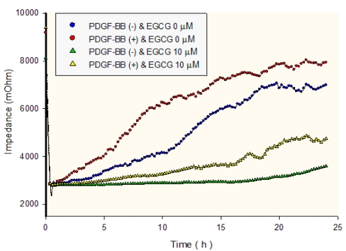
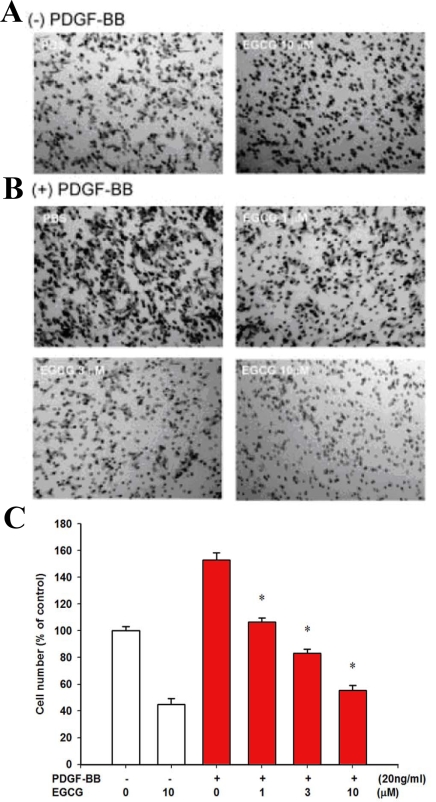
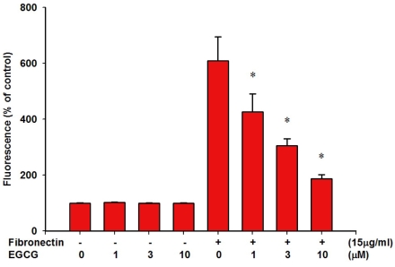
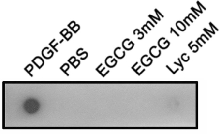
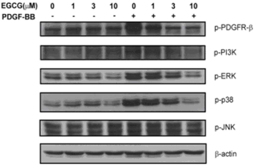
Methods: The migration of RPE cells was detected by an electric cell-substrate impedance sensing (ECIS) migration assay and a Transwell migration assay. Cells were loaded with 2',7'-bis-(carboxyethyl)-5(6')-carboxyfluorescein acetoxymethyl ester (BCECF/AM), and their adhesion to fibronectin was examined. The interactions of EGCG with PDGF-BB were analyzed by a dot binding assay. Cytoskeletal reorganization was examined by immunofluorescence microscopy. The PDGF-BB-induced signaling pathways were detected by western blotting.
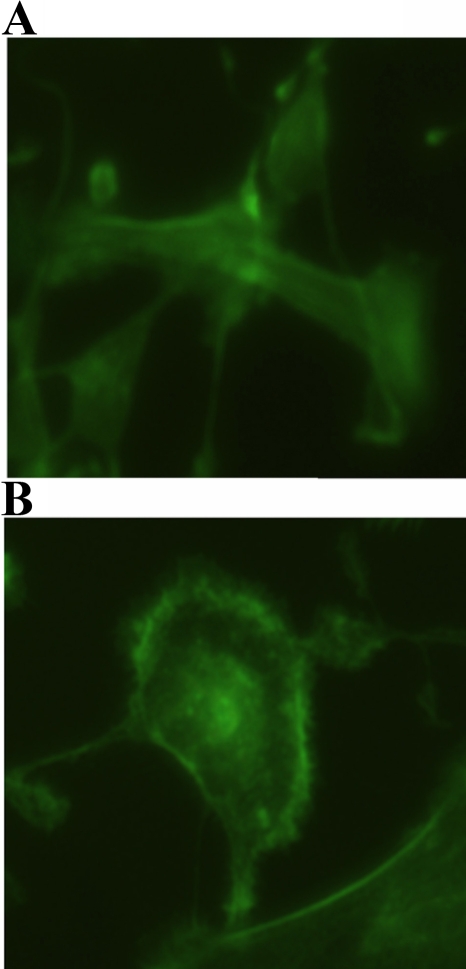
Results: In the present study, we find that EGCG can inhibit PDGF-BB-induced human RPE cell migration and, in a dose-dependent manner, RPE cell adhesion to fibronectin. Our analysis demonstrates that EGCG does not directly bind to PDGF-BB and the inhibition of EGCG against fibronectin-induced cytoskeletal reorganization is observed. Furthermore, EGCG is shown to suppress PDGF-BB-induced PDGF-beta receptors, downstream PI3K/Akt, and MAPK phosphorylation.
Conclusions: Our results provide the first evidence that EGCG is an effective inhibitor of RPE cell migration and adhesion to fibronectin and, therefore, may prevent epiretinal membrane formation.
Figures
Similar articles
-
Inhibitory effects of resveratrol on PDGF-BB-induced retinal pigment epithelial cell migration via PDGFRβ, PI3K/Akt and MAPK pathways.PLoS One. 2013;8(2):e56819. doi: 10.1371/journal.pone.0056819. Epub 2013 Feb 14. PLoS One. 2013. PMID: 23457620 Free PMC article.
-
Crocetin inhibits PDGF-BB-induced proliferation and migration of retinal pigment epithelial cells.Eur J Pharmacol. 2019 Jan 5;842:329-337. doi: 10.1016/j.ejphar.2018.11.001. Epub 2018 Nov 2. Eur J Pharmacol. 2019. PMID: 30395849
-
Lycopene inhibits PDGF-BB-induced retinal pigment epithelial cell migration by suppression of PI3K/Akt and MAPK pathways.Biochem Biophys Res Commun. 2009 Oct 9;388(1):172-6. doi: 10.1016/j.bbrc.2009.07.155. Epub 2009 Aug 5. Biochem Biophys Res Commun. 2009. PMID: 19664603
-
A selective cyclic integrin antagonist blocks the integrin receptors alphavbeta3 and alphavbeta5 and inhibits retinal pigment epithelium cell attachment, migration and invasion.BMC Ophthalmol. 2005 Jun 29;5:16. doi: 10.1186/1471-2415-5-16. BMC Ophthalmol. 2005. PMID: 15987521 Free PMC article.
-
Minireview: Fibronectin in retinal disease.Exp Biol Med (Maywood). 2017 Jan;242(1):1-7. doi: 10.1177/1535370216675245. Epub 2016 Oct 20. Exp Biol Med (Maywood). 2017. PMID: 27798121 Free PMC article. Review.
Cited by
-
DPP-4 inhibitors promote proliferation and migration of rat brain microvascular endothelial cells under hypoxic/high-glucose conditions, potentially through the SIRT1/HIF-1/VEGF pathway.CNS Neurosci Ther. 2019 Mar;25(3):323-332. doi: 10.1111/cns.13042. Epub 2018 Aug 23. CNS Neurosci Ther. 2019. PMID: 30136405 Free PMC article.
-
Epigallocatechin gallate & curcumin prevent transforming growth factor beta 1-induced epithelial to mesenchymal transition in ARPE-19 cells.Indian J Med Res. 2017 Nov;146(Suppl):S85-S96. doi: 10.4103/ijmr.IJMR_1583_15. Indian J Med Res. 2017. PMID: 29578200 Free PMC article.
-
Effects of bradykinin on TGF‑β1‑induced epithelial‑mesenchymal transition in ARPE‑19 cells.Mol Med Rep. 2018 Apr;17(4):5878-5886. doi: 10.3892/mmr.2018.8556. Epub 2018 Feb 2. Mol Med Rep. 2018. PMID: 29436636 Free PMC article.
-
Enhanced delivery of biodegradable mPEG-PLGA-PLL nanoparticles loading Cy3-labelled PDGF-BB siRNA by UTMD to rat retina.J Biosci. 2017 Jun;42(2):299-309. doi: 10.1007/s12038-017-9677-6. J Biosci. 2017. PMID: 28569253
-
Prometastatic NEDD9 Regulates Individual Cell Migration via Caveolin-1-Dependent Trafficking of Integrins.Mol Cancer Res. 2015 Mar;13(3):423-38. doi: 10.1158/1541-7786.MCR-14-0353. Epub 2014 Oct 15. Mol Cancer Res. 2015. PMID: 25319010 Free PMC article.
References
-
- Campochiaro PA. Pathogenic mechanisms in proliferative vitreoretinopathy. Arch Ophthalmol. 1997;115:237–41. - PubMed
-
- Esser P, Heimann K, Bartz-schmidt KU, Fontana A, Schraermeyer U, Thumann G, Weller M. Apoptosis in proliferative vitreoretinal disorders: possible involvement of TGF-beta-induced RPE cell apoptosis. Exp Eye Res. 1997;65:365–78. - PubMed
-
- Miller H, Miller B, Ryan SJ. The role of retinal pigment epithelium in the involution of subretinal neovascularization. Invest Ophthalmol Vis Sci. 1986;27:1644–52. - PubMed
-
- Abe T, Durlu YK, Tamai M. The properties of retinal pigment epithelial cells in proliferative vitreoretinopathy compared with cultured retinal pigment epithelial cells. Exp Eye Res. 1996;63:201–10. - PubMed
-
- Limb GA, Little BC, Meager A, Ogilvie JA, Wolstencroft RA, Franks WA, Chignell AH, Dumonde DC. Cytokines in proliferative vitreoretinopathy. Eye. 1991;5:686–93. - PubMed
Publication types
MeSH terms
Substances
LinkOut - more resources
Full Text Sources
Research Materials
Miscellaneous