

Optical Imaging of Bacterial Infection Models
- PMID: 20376332
- PMCID: PMC2849117
- DOI: 10.1016/j.ddmod.2007.07.001
Optical Imaging of Bacterial Infection Models
Abstract
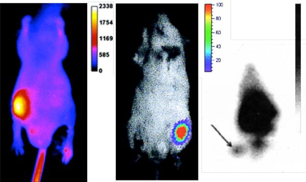
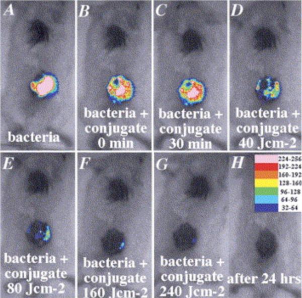
Over the last thirteen years, the field of optical imaging has expanded from in vitro fluorescence microscopy of cells to in vivo imaging of living animals. Recent advances in optical imaging of bacterial infection have been propelled by the invention of genetic methods that produce fluorescent and bioluminescent bacteria, and also the discovery of synthetic fluorescent probes that selectively target bacterial cell surfaces. Optical imaging is an effective method of conducting longitudinal studies of bacterial infection in small animals such as nude mice. It can be used to address questions in medical microbiology concerning migration and colonization and it is an attractive method for determining the efficacy of antibiotic therapies.
Figures


References
-
- Jaffer FA, et al. Molecular Imaging in the Clinical Arena. JAMA. 2005;293:855–862. - PubMed
-
- Hudson M. The Welfare and Scientific Advantages of Non-invasive Imaging of Animals Used in Biomedical Research. Anim. Welfare. 2005;14:303–317.
-
- Doyle TC, et al. In Vivo Bioluminescence Imaging for Integrated Studies of Infection. Cellular Microbiol. 2004;6:303–317. - PubMed
-
- Bashkatov AN, et al. Optical Properties of Human Skin, Subcutaneous and Mucous Tissues in the Wavelength Range from 400 to 2000 nm. J. Phys. D: Appl. Phys. 2005;38:2543–2555.
Grants and funding
LinkOut - more resources
Full Text Sources
Other Literature Sources