. 2010 Apr 1;5(4):e9945.
doi: 10.1371/journal.pone.0009945.

HSPG-binding peptide corresponding to the exon 6a-encoded domain of VEGF inhibits tumor growth by blocking angiogenesis in murine model

Affiliations

HSPG-binding peptide corresponding to the exon 6a-encoded domain of VEGF inhibits tumor growth by blocking angiogenesis in murine model

Tong-Young Lee et al. PLoS One. .

Abstract

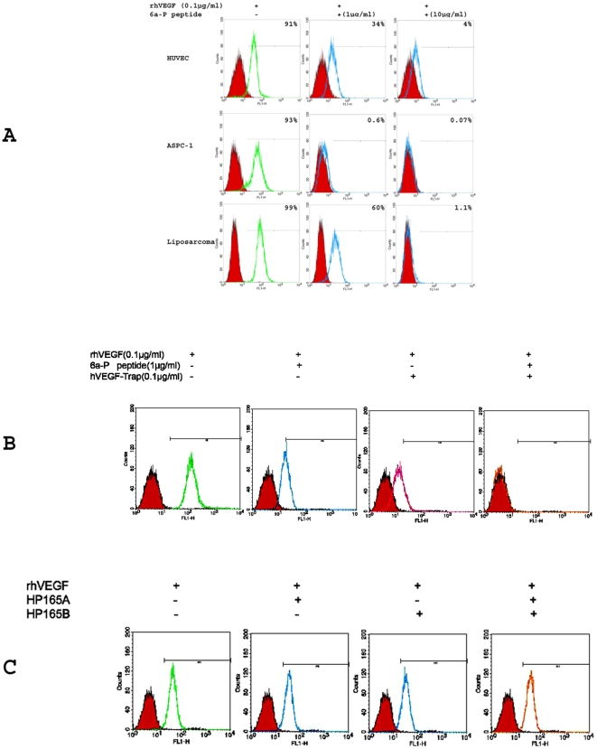
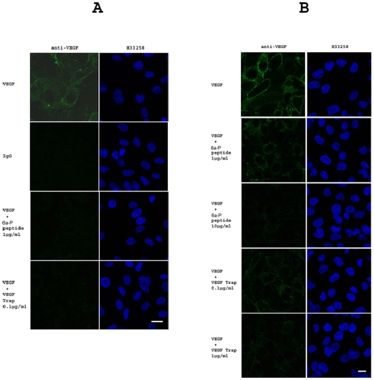
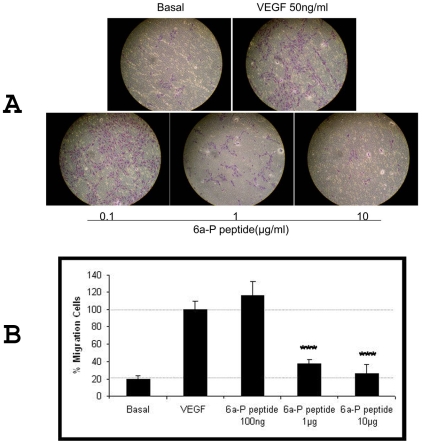
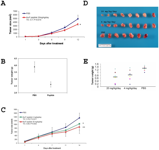
Vascular endothelial growth factor VEGF(165) is a critical element for development of the vascular system in physiological and pathological angiogenesis. VEGF isoforms have different affinities for heparan sulphate proteoglycan (HSPG) as well as for VEGF receptors; HSPGs are important regulators in vascular development. Therefore, inhibition of interactions between VEGF and HSPGs may prevent angiogenesis. Here, we demonstrate that an HSPG-binding synthetic peptide, corresponding to exon 6a-encoded domain of VEGF gene, has anti-angiogenic property. This 20 amino acids synthetic peptide prevents VEGF(165) binding to several different cell types, mouse embryonic sections and inhibits endothelial cell migration, despite its absence in VEGF(165) sequence. Our in vivo anti-tumor studies show that the peptide inhibits tumor growth in both mouse Lewis-Lung Carcinoma and human Liposarcoma tumor-bearing animal models. This is the first evidence that a synthetic VEGF fragment corresponding to exon 6a has functional antagonism both in vitro and in vivo. We conclude that the above HPSG binding peptide (6a-P) is a potent inhibitor of angiogenesis-dependent diseases.


Conflict of interest statement

Competing Interests: The authors have declared that no competing interests exist.

Figures

Figure 1
Figure 1. 6a-P peptide prevents VEGF binding to different cell types by FACS analysis
. HUVECs, fibroblasts, AsPC-1 and liposarcoma cells were subjected to FACS analysis. Cells were digested and resuspensed with 2% BSA in PBS for blocking, 0.1 µg/ml rhVEGF was incubated and present or absent with 1 (low dose) or 10 (high dose) µg/ml of the peptide for 1 h in 4°C. After cold PBS washing, the samples were incubated with VEGF antibody and secondary FITC-labeled antibody. The results show that rhVEGF binding activity is inhibited when the peptide is present (A). VEGF-Trap has been used for a positive control, inhibiting VEGF binding about 70%. The synergic result shows that the rhVEGF binding is inhibited completely when 1 µg/ml of the peptide plus 0.1 µg/ml of VEGF-Trap are present with HUVECs (B). HP165A, HP165B peptides do not inhibit binding of VEGF to HUVECs (C).
Figure 2
Figure 2. 6a-P peptide decreases VEGF binding activity to cell surface by immunocytochemistry.
Immunocytochemistry was used to confirm the FACS results. VEGF (0.1 µg/ml) binding intensity on HUVECS is decreased when the peptide (1 µg/ml) or VEGF-Trap (0.1 µg/ml) are present (A) (Bar, 20 µm). Similar result is shown on human liposarcoma. VEGF intensity is decreased in dose-dependent manner by 6a-P peptide (B) (Bar, 20 µm).
Figure 3
Figure 3. 6a-P peptide inhibits endothelial cells migration.
HUVECs were plated into inserts (8 µm pore size) of 24-well transwell plate and duplicated. The lower chamber was filled with serum free EBM medium containing 50 ng/ml rhVEGF plus different concentrations of the peptide for 16 h. The results show that endothelial cells migrating across the membrane are suppressed by the peptide in dose-dependent manner (A). VEGF induces endothelial cell migration is inhibited almost 90% when peptide is present at high concentration (B; P<0.001).
Figure 4
Figure 4. Angiogenesis and mouse LLC tumor growth are suppressed by 6a-P peptide in vivo.
Two different tumor-bearing animal models were employed for anti-tumor study. The mice were treated with the peptide or PBS twice a day after tumor cells injection. In mouse LLC animal model, the tumor sizes are decreased about 30% after the treatments (n = 5) (A). However, the tumor weights show about 40% different between treated or non-treated groups (B). Human liposarcoma animal model shows the tumor sizes are decreased about 36% in high dose treated group (C). Less hemorrhage (as shows in red on tumor surface) is observed in the high dose of peptide treated despite similarities in tumor sizes for treated and untreated mice (D). The tumor weights show a difference of 2-fold following treatment (n = 8) (E).
Figure 5
Figure 5. Tumor vessel activity is inhibited by 6a-P peptide.
To verify the inhibition of complex of VEGF with KDR, we used the Gv39M monoclonal antibody to recognize VEGF/KDR complex in both peptide and PBS treated xenograft animal sections. Immunohistochemistry result shows that VEGF/KDR complexes are colocalized with vessels (VWF staining) on PBS treated tumor (A; up panel). However, the staining signal is decreased after peptide treatment (A; lower panel). The TUNEL assay shows more apoptotic cells in treated than untreated groups on tumor (B). (Bar, 100 µm)

References

    1. Folkman J. Angiogenesis. Annu Rev Med; 2006;57:1–18. - PubMed
    1. Carmeliet P. Angiogenesis in health and disease. Nat Med. 2003;9:653–60. - PubMed
    1. Tournaire R, Simon1 MP, Noble F, Eichmann A, England P, et al. A short synthetic peptide inhibits signal transduction, migration and angiogenesis mediated by Tie2 receptor. EMBO Rep. 2004;5:262–7. - PMC - PubMed
    1. Robinson JC, Stringer SE. The splice variants of vascular endothelial growth factor (VEGF) and their receptors. J Cell Scien. 2001;114:853–65. - PubMed
    1. Hervě MA, Buteau-Lozanoa H, Mourahb S, Calvob F, Perrot-Applanat M. VEGF189 stimulates endothelial cells proliferation and migration in vitro and up-regulates the expression of Flk-1/KDR mRNA. Exp Cell Res. 2005;309:24–31. - PubMed

Publication types

MeSH terms