. 2010 Apr 1;5(4):e9961.
doi: 10.1371/journal.pone.0009961.

Natural spike trains trigger short- and long-lasting dynamics at hippocampal mossy fiber synapses in rodents

Affiliations

Natural spike trains trigger short- and long-lasting dynamics at hippocampal mossy fiber synapses in rodents

Anja Gundlfinger et al. PLoS One. .

Abstract

Background: Synapses exhibit strikingly different forms of plasticity over a wide range of time scales, from milliseconds to hours. Studies on synaptic plasticity typically use constant-frequency stimulation to activate synapses, whereas in vivo activity of neurons is irregular.

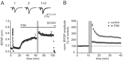
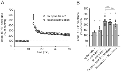
Methodology/principal findings: Using extracellular and whole-cell electrophysiological recordings, we have here studied the synaptic responses at hippocampal mossy fiber synapses in vitro to stimulus patterns obtained from in vivo recordings of place cell firing of dentate gyrus granule cells in behaving rodents. We find that synaptic strength is strongly modulated on short- and long-lasting time scales during the presentation of the natural stimulus trains.

Conclusions/significance: We conclude that dynamic short- and long-term synaptic plasticity at the hippocampal mossy fiber synapse plays a prominent role in normal synaptic function.


Conflict of interest statement

Competing Interests: The authors have declared that no competing interests exist.

Figures

Figure 1
Figure 1. Spiking activity of dentate gyrus granule cells during exploratory behaviour evokes large short-term plasticity at mossy fiber synapses in vitro.
(A) Color-code depicts the location-dependent modulation of firing frequency in Hz of a putative dentate gyrus granule cell while the animal was exploring a rectangular environment. A place field is visible in the down-left corner. (B) Time-resolved plot of spike train 1 (up) of a ∼15 minute recording session of putative granule cell activity, that (A) was based on, and continuous recording of mossy fiber fEPSP (down) in response to presentation of that spike train in vitro. Stimulation artifacts were removed for clarity. Red bars indicate episodes inside the place field. (C) Representative recording of mossy fiber fEPSPs during response to spike train 1, where highly dynamic fEPSP amplitudes were evoked. Grey indicates responses to spikes outside the cell's place field, red indicates responses within. (D) Summary of n = 5 such experiments. Short-term synaptic dynamics were reproducible and comparable between experiments. Data points depict mean ± sem. (E) Pronounced short-term facilitation of fEPSPs is induced by stimulus bursts during traversal of place fields. Line graphs show the first 10 mossy fiber fEPSP response amplitudes during six episodes of place field spiking activity (as indicated by red in C). Red circles indicate responses during 1st burst, white circles responses during 6th burst, redish shadings correspond to bursts in between. Single exemplary recording is shown. (F) Mossy fiber fEPSP amplitudes decrease again during the first 10 stimuli after place field traversal indicated by grey shadings. Single exemplary recording is shown. (G/H) Field EPSP response amplitude as a function of preceding inter-stimulus interval during spike train 1. A large dynamic range of response amplitudes is apparent with amplitudes generally largest with ISIs ranging from 50–1000 ms. (G) depicts single exemplary recording, (H) summarizes n = 6 such experiments. Red circles indicate fEPSP responses during spikes of place field traversal, grey circles such outside of place fields. Blue dashed lines in panels C to H indicate basal response amplitudes to constant stimulation at 0.05 Hz.
Figure 2
Figure 2. Place field specific spiking activity of dentate gyrus granule cells triggers long-term potentiation of mossy fiber synaptic responses in vitro.
(A) Presentation of spike train 1 (indicated by grey area) led to potentiation of mossy fiber fEPSP amplitudes in this examplary recording. Constant stimulation frequency before and after delivery of spike train was 0.05 Hz. Application of DCGIV at the end of experiment blocked mossy fiber synaptic transmission. Upper traces show averages of 10 sweeps under control condition and 30 min after presentation of spike train 1. (B) Summary of n = 5 such experiments. Presentation of spike train led to reliable long-term potentiation of fEPSP amplitudes to ∼130% of control values 25 min after spike train 1. (C) Time-resolved plot of another place field specific spike episode (spike train 2, up) and continuous recording of mossy fiber fEPSP response to single presentation of this spike train (lower part). Stimulus artifacts are cut for visual clarity. Please note different timescale compared to spike train 1. (D) Examplary mossy fiber synaptic fEPSP recording, where a single presentation of spike train 2 (grey bar, not drawn to scale) leads to long-term potentiation of fEPSP responses. Arrow points to frequency facilitation paradigm (switch of stimulation frequency from 0.05 Hz to 1 Hz for 20 stimuli). Application of DCGIV (1 µM) at the end of experiment blocked mossy fiber fEPSPs. Upper traces show averages of 10 sweeps each under control condition and 25 minutes after presentation of spike train. Constant stimulation frequency was 0.05 Hz. (E) Repetitive presentation of spike train 2 (5x with 30 s pauses inbetween) resulted in pronounced long-term potentiation of mossy fiber fEPSP amplitudes in this examplary experiment. Upper traces show averages of 10 sweeps each under control condition and 25 minutes after repetitive presentation of spike train. (F) Summary of n = 6 experiments with single presentation of spike train (open circles) and n = 7 experiments with repetitive presentation (filled circles). Both paradigms led to significant long-term potentiation of response amplitudes to ∼150% and ∼230% of control values, respectively. Data shows mean ± sem. Upper dashed lines in subpanels indicate basal response amplitudes to constant stimulation at 0.05 Hz.
Figure 3
Figure 3. Natural spike trains induce mossy fiber LTP indepedent of NMDAR and mGluR activation.
(A) Exemplary mossy fiber fEPSP recording under blockage of NMDA- and mGlu-receptors. Repetitive presentation of spike train 2 still induced significant long-term potentiation. Arrow points to frequency facilitation paradigm. Upper traces are averages of 10 sweeps each under control condition and 25 min after presentation of spike trains. Constant stimulation frequency was 0.05 Hz. (B) Summary of n = 6 such experiments and experiments under control conditions, respectively. Repetitive presentation of spike train 2 resulted in potentiation of response amplitudes to ∼220% of control values 30 min after presentation of spike trains. Data was binned to 1 min time points and depicts mean ± sem. Upper dashed lines in subpanels indicate basal response amplitudes to constant stimulation at 0.05 Hz.
Figure 4
Figure 4. Mossy fiber synaptic LTP - induced by place field specific spiking activity of dentate gyrus granule cells - is presynaptically expressed.
(A) Examplary whole-cell recording of mossy fiber synaptic responses in CA3 pyramidal cell. Repetitive presentation of spike train 2 (grey bars, compare Figure 2) induces long-term potentiation of EPSC amplitudes. Upper traces show averages of 10 sweeps each under control condition and 20 min after presentation of spike train. Constant stimulation frequency outside of spike train 2 was 0.1 Hz. CA3 pyramidal cell was held in voltage-clamp condition at -60 mV, also during presentation of spike train. Upper dashed line indicates basal response amplitudes to constant stimulation at 0.1 Hz. (B) Summary of n = 5 whole-cell experiments where repetitive presentation of spike train 2 induces long-term potentiation of mossy fiber EPSC amplitudes. Potentiation to ∼220% of control values was visible 30 min after spike train. Data was binned to 0.5 min time points and depicts mean ± sem. (C) CV2 analysis of data from experiments in A. The change in the squared coefficient of variation in control versus LTP condition shows a linear dependence on the change in the mean response amplitude. (D) The mean rate of failures of synaptic transmission is decreased after expression of LTP. Upper traces show 50 individual sweeps (grey) and mean sweeps (black) in control and LTP condition of an exemplary whole-cell recording. Note the large incidence of synaptic failures under control conditions.
Figure 5
Figure 5. Natural spike train induced long-term potentiation is strongly reduced in the presence of elevated cAMP levels.
(A) Application of the adenylate cyclase activator forskolin (50 µM) enhances synaptic transmission in this exemplary experiment and strongly reduces long-term potentiation induced by repetitive (5 x) delivery of spike train 2. Traces on top are averages of five consecutive sweeps taken at the time point indicated by the numbers in the graph. Triangle denotes frequency facilitation paradigm for 20 pulses with 1 Hz, arrow indicates time point of spike train 2 application, second horizontal bar represents application of DCGIV (1 µM) at the end of experiment. (B) Summary plot displaying the drastically reduced potentiation for n = 4 such experiments (closed circles). Values are normalized to the amplitude in forskolin before train delivery. For comparison, the potentiation elicited by spike train 2 in the absence of drugs (open circles, same dataset as in Figure 2F) is overlayed. In the presence of forskolin the potentiation was reduced to 143.4±11.5% (p<0.001, compared to control).
Figure 6
Figure 6. Summary of the amount of potentiation induced by different stimulation paradigms.
(A) Long-term potentiation induced by repetitive presentation of spike train 2 (open circles) is comparable to LTP induced by a classical LTP induction protocol (grey circles) by tetanic stimulation (3×125 pulses at 25 Hz with 30 s pauses in between). (B) All used stimulation paradigms based on place field specific spiking activity of granule cells triggered a significant long-term potentiation of synaptic responses when compared to control values. Asterisks point to a significant increase in fEPSP amplitude (single distribution t-test against 100%). The amount of LTP induced by repetitive presentation of spike train 2 was not significantly (ns) different from LTP induced by a classical tetanic paradigm or under the action of AP-V and LY341496.

Similar articles

Cited by

References

    1. Malenka RC, Nicoll RA. Long-term potentiation–a decade of progress? Science. 1999;285:1870–1874. - PubMed
    1. Zucker RS, Regehr WG. Short-term synaptic plasticity. Annu Rev Physiol. 2002;64:355–405. - PubMed
    1. O'Keefe J. A review of the hippocampal place cells. Prog Neurobiol. 1979;13:419–439. - PubMed
    1. O'Keefe J, Dostrovsky J. The hippocampus as a spatial map. Preliminary evidence from unit activity in the freely-moving rat. Brain Res. 1971;34:171–175. - PubMed
    1. Wilson MA, McNaughton BL. Dynamics of the hippocampal ensemble code for space. Science. 1993;261:1055–1058. - PubMed

Publication types