

Robustness of the retinotopic attentional trace after eye movements
- PMID: 20377296
- PMCID: PMC3213860
- DOI: 10.1167/10.3.19
Robustness of the retinotopic attentional trace after eye movements
Abstract
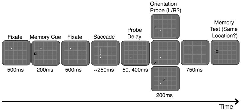
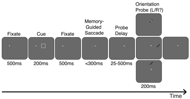
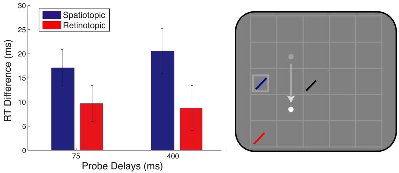
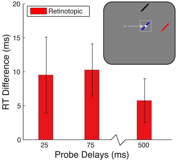
With each eye movement, the image received by the visual system changes drastically. To maintain stable spatiotopic (world-centered) representations, the relevant retinotopic (eye-centered) coordinates must be continually updated. Although updating or remapping of visual scene representations can occur very rapidly, J. D. Golomb, M. M. Chun, and J. A. Mazer (2008) demonstrated that representations of sustained attention update more slowly than the remapping literature would predict; attentional benefits at previously attended retinotopic locations linger after completion of the saccade, even when this location is no longer behaviorally relevant. The present study explores the robustness of this "retinotopic attentional trace." We report significant retinotopic facilitation despite attempts to eliminate or reduce it by enhancing spatiotopic reference frames with permanent visual cues in the stimulus display and by introducing a different task where the attended location is the saccade target itself. Our results support and extend our earlier model of native retinotopically organized salience maps that must be dynamically updated to reflect the task-relevant spatiotopic location with each saccade. Consistent with the idea that attentional facilitation arises from persistent, recurrent neural activity, it takes measurable time for this facilitation to decay, leaving behind a retinotopic attentional trace after the saccade has been executed, regardless of conflicting task demands.
Figures
Similar articles
-
Visual Remapping.Annu Rev Vis Sci. 2021 Sep 15;7:257-277. doi: 10.1146/annurev-vision-032321-100012. Epub 2021 Jul 9. Annu Rev Vis Sci. 2021. PMID: 34242055 Free PMC article. Review.
-
The native coordinate system of spatial attention is retinotopic.J Neurosci. 2008 Oct 15;28(42):10654-62. doi: 10.1523/JNEUROSCI.2525-08.2008. J Neurosci. 2008. PMID: 18923041 Free PMC article.
-
Attentional facilitation throughout human visual cortex lingers in retinotopic coordinates after eye movements.J Neurosci. 2010 Aug 4;30(31):10493-506. doi: 10.1523/JNEUROSCI.1546-10.2010. J Neurosci. 2010. PMID: 20685992 Free PMC article.
-
Attention doesn't slide: spatiotopic updating after eye movements instantiates a new, discrete attentional locus.Atten Percept Psychophys. 2011 Jan;73(1):7-14. doi: 10.3758/s13414-010-0016-3. Atten Percept Psychophys. 2011. PMID: 21258903 Free PMC article.
-
Constructing stable spatial maps of the world.Perception. 2012;41(11):1355-72. doi: 10.1068/p7392. Perception. 2012. PMID: 23513621 Review.
Cited by
-
The coordinate system of endogenous spatial attention during smooth pursuit.J Vis. 2020 Jul 1;20(7):26. doi: 10.1167/jov.20.7.26. J Vis. 2020. PMID: 32720972 Free PMC article.
-
Decoding Remapped Spatial Information in the Peri-Saccadic Period.J Neurosci. 2024 Jul 24;44(30):e2134232024. doi: 10.1523/JNEUROSCI.2134-23.2024. J Neurosci. 2024. PMID: 38871460 Free PMC article.
-
Visual Remapping.Annu Rev Vis Sci. 2021 Sep 15;7:257-277. doi: 10.1146/annurev-vision-032321-100012. Epub 2021 Jul 9. Annu Rev Vis Sci. 2021. PMID: 34242055 Free PMC article. Review.
-
Not all attention orienting is created equal: recognition memory is enhanced when attention orienting involves distractor suppression.Neurobiol Learn Mem. 2015 Apr;120:28-40. doi: 10.1016/j.nlm.2015.02.006. Epub 2015 Feb 17. Neurobiol Learn Mem. 2015. PMID: 25701278 Free PMC article.
-
Rapid updating of spatial working memory across saccades.Sci Rep. 2018 Jan 18;8(1):1072. doi: 10.1038/s41598-017-18779-9. Sci Rep. 2018. PMID: 29348583 Free PMC article.
References
-
- Abrams RA, Pratt J. Oculocentric coding of inhibited eye movements to recently attended locations. Journal of Experimental Psychology: Human Perception and Performance. 2000;26:776–788. - PubMed
-
- Afraz A, Cavanagh P. The gender-specific face aftereffect is based in retinotopic not spatiotopic coordinates across several natural image transformations. Journal of Vision. 2009. pp. 10pp. 1–17. http://journalofvision.org/9/10/10/ - DOI - PMC - PubMed
-
- Awh E, Armstrong KM, Moore T. Visual and oculomotor selection: Links, causes and implications for spatial attention. Trends in Cognitive Sciences. 2006;10:124–130. - PubMed
-
- Bellebaum C, Hoffmann KP, Daum I. Post-saccadic updating of visual space in the posterior parietal cortex in humans. Behavioural Brain Research. 2005;163:194–203. - PubMed
-
- Bisley JW, Goldberg ME. Neuronal activity in the lateral intraparietal area and spatial attention. Science. 2003;299:81–86. - PubMed
Publication types
MeSH terms
Grants and funding
LinkOut - more resources
Full Text Sources
