

Three-dimensional structure of recombinant type 1 inositol 1,4,5-trisphosphate receptor
- PMID: 20377523
- PMCID: PMC3685215
- DOI: 10.1042/BJ20100143
Three-dimensional structure of recombinant type 1 inositol 1,4,5-trisphosphate receptor
Abstract
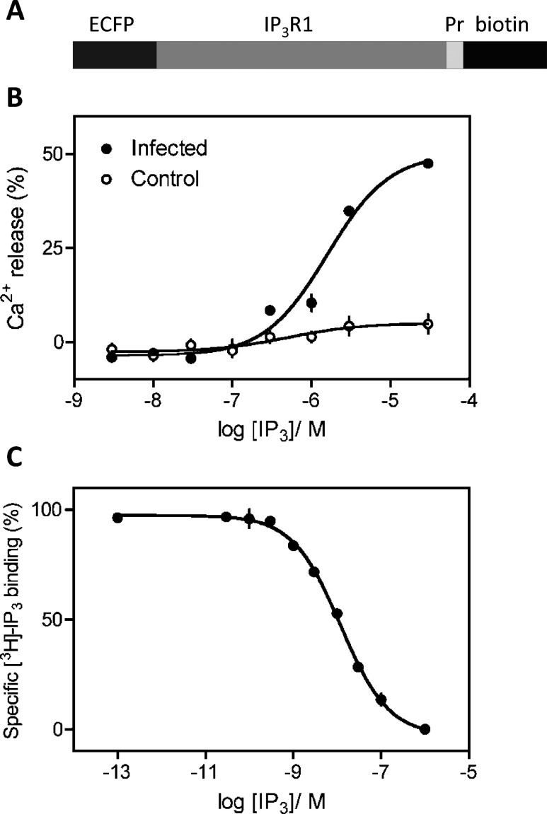
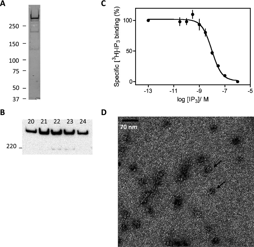
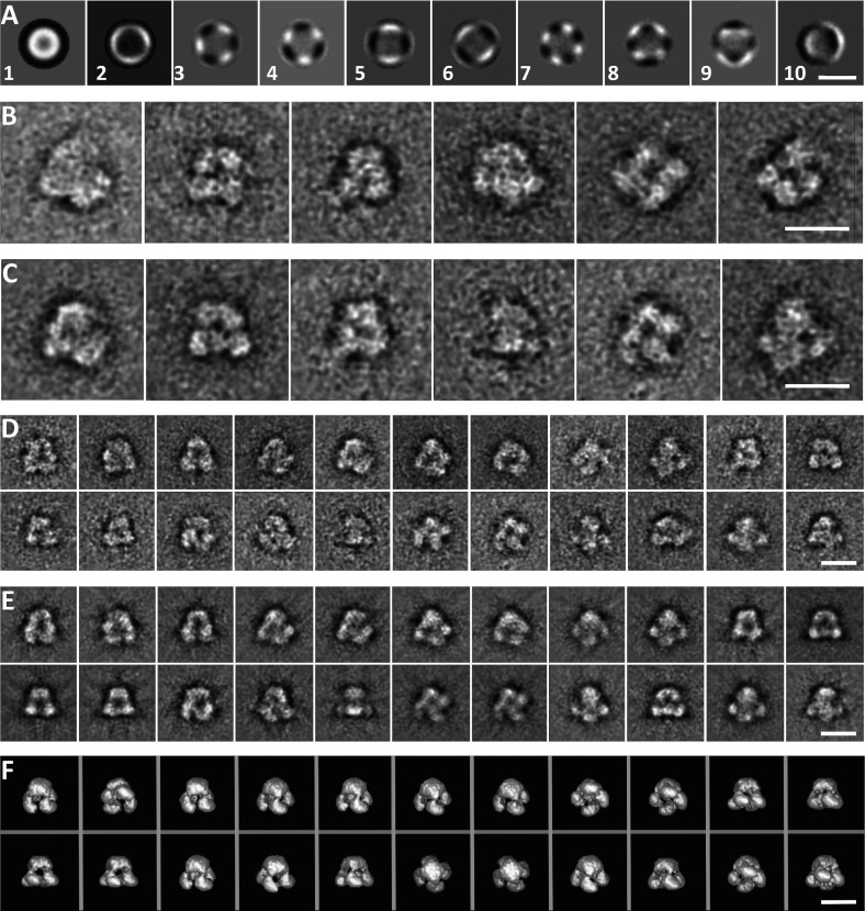
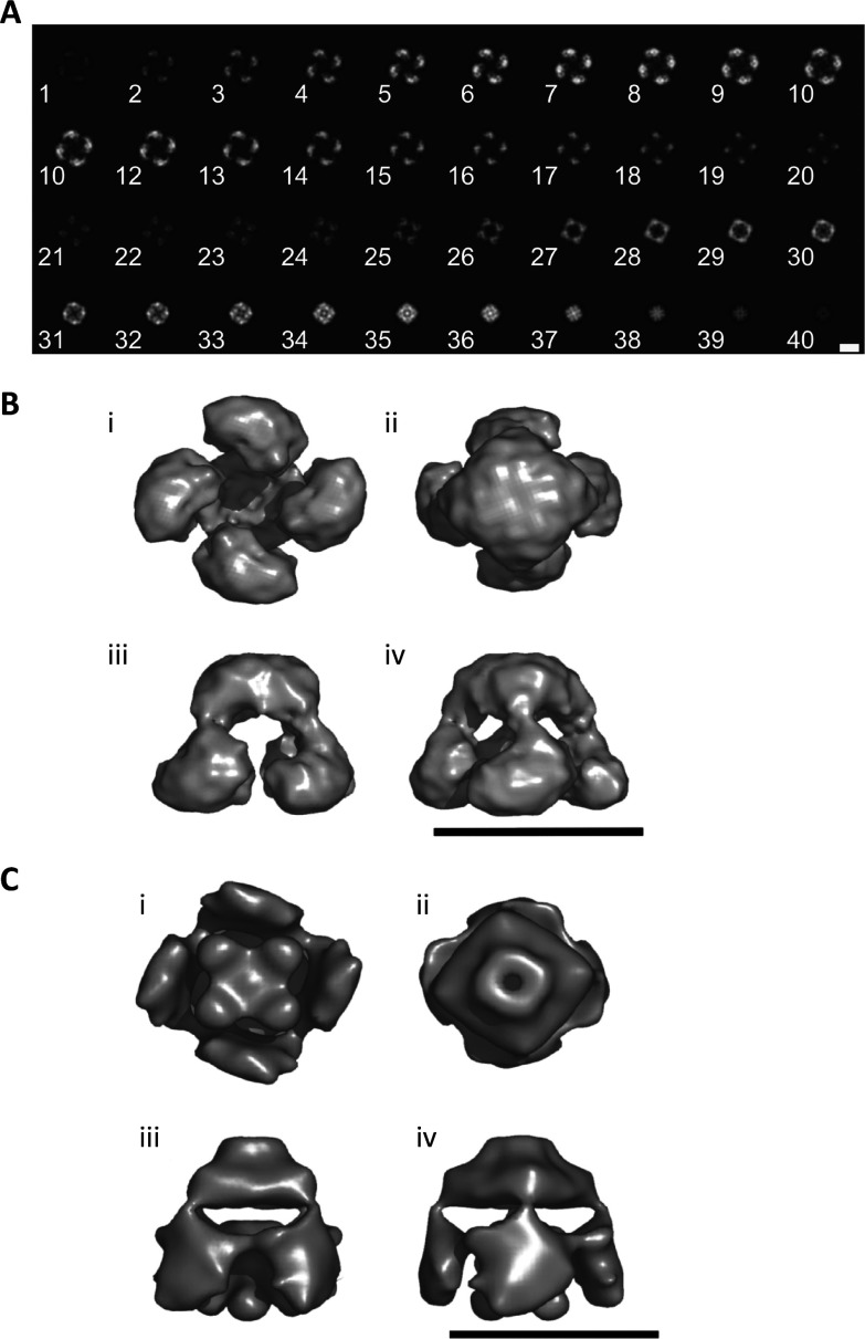
IP3Rs (inositol 1,4,5-trisphosphate receptors) are the intracellular channels that mediate release of Ca2+ from the endoplasmic reticulum in response to the many stimuli that evoke Ins(1,4,5)P3 formation. We characterized and purified type 1 IP3R heterologously expressed in Sf9 insect cells, and used the purified IP3R1 to determine its three-dimensional structure by electron microscopy and single-particle analysis. Recombinant IP3R1 has 4-fold symmetry with overall dimensions of approx. 19.5 nm x 19.5 nm x 17.5 nm. It comprises a small domain, which is likely to include the pore, linked by slender bridges to a large cytoplasmic domain with four petal-like regions. Our structures of recombinant IP3R1 and native cerebellar IP3R have similar appearances and dimensions. The only notable difference is the absence of a central stigma-like domain from the cytoplasmic region of recombinant IP3R1. The first structure of a recombinant IP3R is an important step towards developing three-dimensional structures of IP3R that better contribute to our understanding of the structural basis of IP3R activation.
Figures
Similar articles
-
Three-dimensional rearrangements within inositol 1,4,5-trisphosphate receptor by calcium.J Biol Chem. 2003 Dec 26;278(52):52881-9. doi: 10.1074/jbc.M309743200. Epub 2003 Oct 30. J Biol Chem. 2003. PMID: 14593123
-
Region-specific proteolysis differentially regulates type 1 inositol 1,4,5-trisphosphate receptor activity.J Biol Chem. 2017 Jul 14;292(28):11714-11726. doi: 10.1074/jbc.M117.789917. Epub 2017 May 19. J Biol Chem. 2017. PMID: 28526746 Free PMC article.
-
80K-H interacts with inositol 1,4,5-trisphosphate (IP3) receptors and regulates IP3-induced calcium release activity.J Biol Chem. 2009 Jan 2;284(1):372-380. doi: 10.1074/jbc.M805828200. Epub 2008 Nov 6. J Biol Chem. 2009. PMID: 18990696
-
The IP3 receptor/Ca2+ channel and its cellular function.Biochem Soc Symp. 2007;(74):9-22. doi: 10.1042/BSS0740009. Biochem Soc Symp. 2007. PMID: 17233576 Review.
-
Toward a high-resolution structure of IP₃R channel.Cell Calcium. 2014 Sep;56(3):125-32. doi: 10.1016/j.ceca.2014.08.002. Epub 2014 Aug 10. Cell Calcium. 2014. PMID: 25159857 Free PMC article. Review.
Cited by
-
EI24 promotes cell adaption to ER stress by coordinating IRE1 signaling and calcium homeostasis.EMBO Rep. 2022 Feb 3;23(3):e51679. doi: 10.15252/embr.202051679. Epub 2022 Jan 10. EMBO Rep. 2022. PMID: 35005829 Free PMC article.
-
Endoplasmic Reticulum-Mitochondrial Contactology: Structure and Signaling Functions.Trends Cell Biol. 2018 Jul;28(7):523-540. doi: 10.1016/j.tcb.2018.02.009. Epub 2018 Mar 24. Trends Cell Biol. 2018. PMID: 29588129 Free PMC article. Review.
-
IP(3) receptors: toward understanding their activation.Cold Spring Harb Perspect Biol. 2010 Dec;2(12):a004010. doi: 10.1101/cshperspect.a004010. Epub 2010 Oct 27. Cold Spring Harb Perspect Biol. 2010. PMID: 20980441 Free PMC article. Review.
-
Permeant calcium ion feed-through regulation of single inositol 1,4,5-trisphosphate receptor channel gating.J Gen Physiol. 2012 Dec;140(6):697-716. doi: 10.1085/jgp.201210804. Epub 2012 Nov 12. J Gen Physiol. 2012. PMID: 23148262 Free PMC article.
-
Inositol (1,4,5)-trisphosphate receptor microarchitecture shapes Ca2+ puff kinetics.Biophys J. 2011 Feb 16;100(4):822-31. doi: 10.1016/j.bpj.2011.01.003. Biophys J. 2011. PMID: 21320425 Free PMC article.
References
-
- Taylor C. W., Genazzani A. A., Morris S. A. Expression of inositol trisphosphate receptors. Cell Calcium. 1999;26:237–251. - PubMed
-
- Dellis O., Dedos S., Tovey S. C., Taufiq-Ur-Rahman Dubel S. J., Taylor C. W. Ca2+ entry through plasma membrane IP3 receptors. Science. 2006;313:229–233. - PubMed
-
- Stehno-Bittel L., Lückhoff A., Clapham D. E. Calcium release from the nucleus by InsP3 receptor channels. Neuron. 1995;14:163–167. - PubMed
Publication types
MeSH terms
Substances
Grants and funding
LinkOut - more resources
Full Text Sources
Miscellaneous