. 2010 Apr 8:10:103.
doi: 10.1186/1471-2180-10-103.

Microarray studies on effects of Pneumocystis carinii infection on global gene expression in alveolar macrophages

Affiliations

Microarray studies on effects of Pneumocystis carinii infection on global gene expression in alveolar macrophages

Bi-Hua Cheng et al. BMC Microbiol. .

Abstract

Background: Pneumocystis pneumonia is a common opportunistic disease in AIDS patients. The alveolar macrophage is an important effector cell in the clearance of Pneumocystis organisms by phagocytosis. However, both the number and phagocytic activity of alveolar macrophages are decreased in Pneumocystis infected hosts. To understand how Pneumocystis inactivates alveolar macrophages, Affymetrix GeneChip RG-U34A DNA microarrays were used to study the difference in global gene expression in alveolar macrophages from uninfected and Pneumocystis carinii-infected Sprague-Dawley rats.

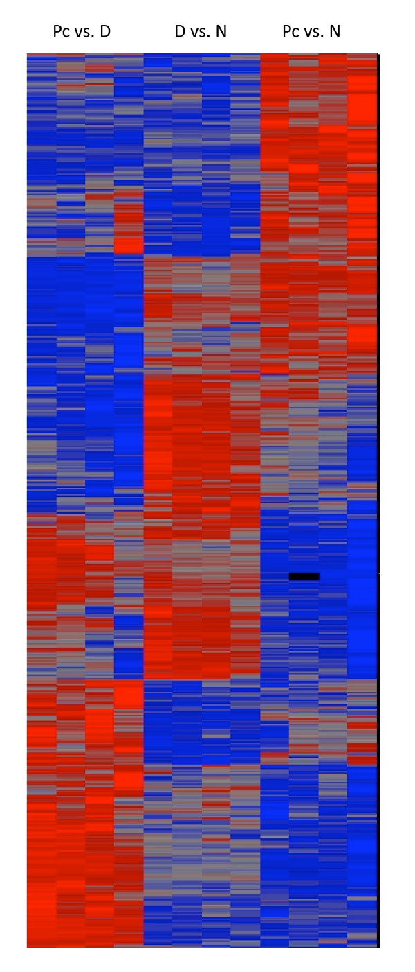
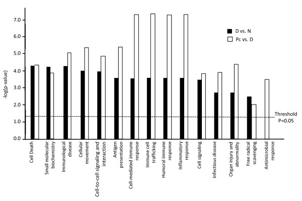
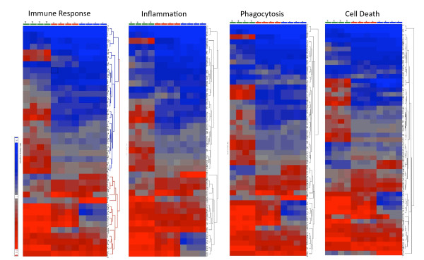
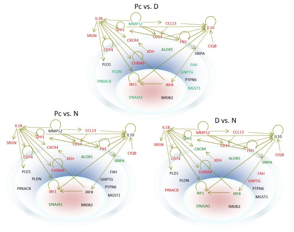
Results: Analyses of genes that were affected by Pneumocystis infection showed that many functions in the cells were affected. Antigen presentation, cell-mediated immune response, humoral immune response, and inflammatory response were most severely affected, followed by cellular movement, immune cell trafficking, immunological disease, cell-to-cell signaling and interaction, cell death, organ injury and abnormality, cell signaling, infectious disease, small molecular biochemistry, antimicrobial response, and free radical scavenging. Since rats must be immunosuppressed in order to develop Pneumocystis infection, alveolar macrophages from four rats of the same sex and age that were treated with dexamethasone for the entire eight weeks of the study period were also examined. With a filter of false-discovery rate less than 0.1 and fold change greater than 1.5, 200 genes were found to be up-regulated, and 144 genes were down-regulated by dexamethasone treatment. During Pneumocystis pneumonia, 115 genes were found to be up- and 137 were down-regulated with the same filtering criteria. The top ten genes up-regulated by Pneumocystis infection were Cxcl10, Spp1, S100A9, Rsad2, S100A8, Nos2, RT1-Bb, Lcn2, RT1-Db1, and Srgn with fold changes ranging between 12.33 and 5.34; and the top ten down-regulated ones were Lgals1, Psat1, Tbc1d23, Gsta1, Car5b, Xrcc5, Pdlim1, Alcam, Cidea, and Pkib with fold changes ranging between -4.24 and -2.25.

Conclusions: In order to survive in the host, Pneumocystis organisms change the expression profile of alveolar macrophages. Results of this study revealed that Pneumocystis infection affects many cellular functions leading to reduced number and activity of alveolar macrophages during Pneumocystis pneumonia.


Figures

Figure 1
Figure 1
Principle component analysis of microarray results. The blue, red, and green oval dots represent linear combinations of the expression data, including relative expression value and variance, of the 8799 genes in AMs from each Normal, Dex, or Dex-Pc rat. The principle component analysis (PCA) software examined three components of genes in different samples for those with similar or different expression profiles. The first component, shown in the x-axis, includes genes with a high degree of variance. The second component, displayed in the y-axis, encompasses genes that had a median range of variance. The third component, represented by z-axis, contains those with a minor variance.
Figure 2
Figure 2
Hierarchical clustering of differentially expressed genes. ANOVA was first performed to identify genes that are differentially expressed due to dexamethasone treatment or Pneumocystis infection. Each lane represents the expression profile of AMs from one rat. The first four lanes show the expression profiles of AMs from the four Dex-Pc rats compared to that of Dex rats, the middle four lanes display those of the four Dex rats compared to that of Normal rats, and the remaining four lanes represent those of the four Dex-Pc rats compared to that of Normal rats. Red and blue colors indicate high and low expression levels, respectively. Gray color indicates no change in expression levels.
Figure 3
Figure 3
Functions affected by dexamethasone or Pneumocystis infection. Cellular functions identified by IPA as being affected by dexamethasone or Pneumocystis infection are illustrated with bar graphs based on the levels of -log(p-value), the higher the levels the more significant of the effect. Black bars indicate functions affected by dexamethasone treatment, while open bars denote those affected by Pneumocystis infection.
Figure 4
Figure 4
Hierarchical clustering of differentially expressed genes related to the major functions of AMs. Genes involved in immune response, inflammation, phagocytosis, and cell death were analyzed. Each lane represents the expression profile of AMs from one rat. For each panel, the first four lanes show the expression profiles of AMs from the four Dex-Pc rats compared to that of Dex rats, the middle four lanes display those of the four Dex rats compared to that of Normal rats, and the remaining four lanes represent those of the four Dex-Pc rats compared to that of Normal rats. Red and blue colors indicate high and low expression levels, respectively. Gray color indicates no change in expression levels.
Figure 5
Figure 5
Subcellular localization of the products of differentially expressed genes during dexamethasone treatment or Pneumocystis infection. The outer ring represents the cell membrane, and the inner oval circle denotes the nucleus; the space between these two structures is the cytoplasm. Locations of the gene products are as indicated. Genes are shown in different colors, with red representing up-regulation and green down-regulation. Genes that have a direct relationship between each other are connected by solid arrows, and those with indirect relationships are linked by dotted arrows.

Similar articles

Cited by

References

    1. Sepkowitz KA. Opportunistic infections in patients with and patients without Acquired Immunodeficiency Syndrome. Clin Infect Dis. 2002;34(8):1098–1107. doi: 10.1086/339548. - DOI - PubMed
    1. Tellez I, Barragán M, Franco-Paredes C, Petraro P, Nelson K, Del Rio C. Pneumocystis jiroveci pneumonia in patients with AIDS in the inner city: a persistent and deadly opportunistic infection. Am J Med Sci. 2008;335(3):192–197. doi: 10.1097/MAJ.0b013e318152004b. - DOI - PubMed
    1. Mocroft A, Sabin CA, Youle M, Madge S, Tyrer M, Devereux H, Deayton J, Dykhoff A, Lipman MC, Phillips AN. Changes in AIDS-defining illnesses in a London Clinic, 1987-1998. J Acquir Immune Defic Syndr. 1999;21(5):401–407. doi: 10.1097/01.pas.0000213299.11649.106. - DOI - PubMed
    1. Matsumoto Y, Matsuda S, Tegoshi T. Yeast glucan in the cyst wall of Pneumocystis carinii . J Protozool. 1989;36(1):21S–22S. - PubMed
    1. Walker AN, Garner RE, Horst MN. Immunocytochemical detection of chitin in Pneumocystis carinii . Infect Immun. 1990;58(2):412–415. - PMC - PubMed

Publication types

MeSH terms