

Case Reports
doi: 10.1177/159101991001600113.
Epub 2010 Mar 25.
Traumatic persistent trigeminal artery--cavernous sinus fistula treated by transcatheter arterial embolization. A case report
Affiliations
- PMID: 20377986
- PMCID: PMC3277963
- DOI: 10.1177/159101991001600113
Item in Clipboard
Case Reports
Traumatic persistent trigeminal artery--cavernous sinus fistula treated by transcatheter arterial embolization. A case report
Interv Neuroradiol.
2010 Mar.
Erratum in
- Interv Neuroradiol. 2010 Jun;16(2):214
Abstract
We describe a rare case of traumatic persistent trigeminal artery (PTA) - cavernous sinus fistula. Cerebral angiography showed direct communication between the right PTA and the cavernous sinus which was treated by transcathether arterial embolization. Although previous reports have indicated the use of more coils to treat this condition, we successfully treated the patient with only two coils placed near the orifice of the fistula after sufficient anatomical evaluation.
Figures
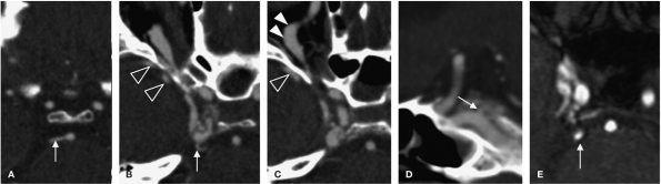
Axial (A-C) and sagittal (D) views of computed tomography angiography. Axial view of magnetic resonance angiography (E). A-C) The right cavernous sinus is distended. Dilated superior ophthalmic vein (full arrowheads) and sphenoparietal sinus (empty arrowheads) are visualized, indicating drainage veins. A,D) The origin and cisternal portion of the persistent trigeminal artery are indicated (arrows). E) The cisternal portion of the right PTA is indicated by an arrow. The origin of the PTA and fistulous point is obscured.
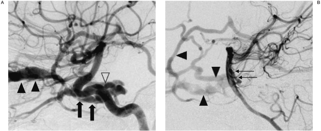
A) Right internal carotid arteriogram, the cavernous sinus (arrows) and dilated SOV (full arrowheads) are seen in a lateral view, revealing the origin of the PTA (empty arrowhead). B) Left vertebral arteriogram, lateral view, showing the cisternal portion of the PTA (arrows) filling the fistula, indicating the PTA-cavernous sinus fistula. The cavernous sinus and dilated cortical veins (arrowheads) are also shown.
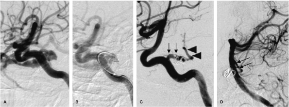
A,B) Lateral oblique view during transarterial embolization. B) A microcatheter was inserted into the fistula, and the two GDC vortex coils were deployed to occlude the fistula. C) Right internal carotid arteriogram after coiling reveals a complete occlusion of the fistula. There is a connection between the preserved PTA (arrows) and the basilar artery (arrowheads). D) The left vertebral arteriogram shows filling of the PTA (arrows).
Similar articles
-
Bilateral persistent trigeminal arteries with unilateral trigeminal artery to cavernous sinus fistula. A case report.Interv Neuroradiol. 2013 Sep;19(3):339-43. doi: 10.1177/159101991301900311. Epub 2013 Sep 26. Interv Neuroradiol. 2013. PMID: 24070083 Free PMC article.
-
Staged Endovascular Occlusion of a Posterior Communicating Artery-Cavernous Sinus Fistula and a Basilar Artery-Cavernous Sinus Fistula Associated with Traumatic Pseudoaneurysms: Technical Consideration and Literature Review.World Neurosurg. 2017 Nov;107:1051.e7-1051.e11. doi: 10.1016/j.wneu.2017.08.070. Epub 2017 Aug 24. World Neurosurg. 2017. PMID: 28842235 Review.
-
Spontaneous Persistent Primitive Trigeminal Artery-Cavernous Sinus Fistula Successfully Treated by Multipronged Coil Embolization: Case Report and Literature Review.World Neurosurg. 2019 Aug;128:122-126. doi: 10.1016/j.wneu.2019.05.003. Epub 2019 May 10. World Neurosurg. 2019. PMID: 31078800 Review.
-
Cavernous sinus fistula caused by intracavernous rupture of a persistent primitive trigeminal artery. Case report.J Neurosurg. 1984 Aug;61(2):391-5. doi: 10.3171/jns.1984.61.2.0391. J Neurosurg. 1984. PMID: 6737065
-
Ruptured Persistent Trigeminal Artery Causing Direct Cavernous Sinus Fistula Treated with Pipeline Embolization and Minimal Coiling.World Neurosurg. 2018 Jan;109:471-475.e1. doi: 10.1016/j.wneu.2017.10.017. Epub 2017 Oct 16. World Neurosurg. 2018. PMID: 29042328
Cited by
-
Persistent trigeminal artery and its variants on MR angiography.Surg Radiol Anat. 2012 Apr;34(3):271-6. doi: 10.1007/s00276-011-0848-0. Epub 2011 Jul 8. Surg Radiol Anat. 2012. PMID: 21739246
-
Clinical Importance of the Persistent Primitive Trigeminal Artery in Vascular Lesions and Its Role in Endovascular Treatment.Front Neurol. 2022 Jul 11;13:928608. doi: 10.3389/fneur.2022.928608. eCollection 2022. Front Neurol. 2022. PMID: 35899260 Free PMC article. Review.
-
Bridging the Gap between Ophthalmology and Emergency Medicine in Community-Based Emergency Departments (EDs): A Neuro-Ophthalmology Guide for ED Practitioners.Clin Pract. 2021 Dec 2;11(4):919-932. doi: 10.3390/clinpract11040106. Clin Pract. 2021. PMID: 34940005 Free PMC article.
-
Anatomical features and clinical relevance of a persistent trigeminal artery.Surg Neurol Int. 2012;3:111. doi: 10.4103/2152-7806.101798. Epub 2012 Sep 28. Surg Neurol Int. 2012. PMID: 23087827 Free PMC article.
References
-
- Geibprasert S, Jiarakonqmun P, et al. Trigeminal fistula treated by combined transvenous and transarterial embolisation. Acta Neurochir (Wien) 2008;150:583–588. - PubMed
-
- Yang X, Mu S, et al. Treatment of trauma tictrigeminal cavernous fistula by coil embolization and compression of carotid artery. Neurol India. 2007;55:396–398. - PubMed
-
- Hahnel S, Hartmann M, et al. Persistent hypoglossal artery: MRI, MRA and digital subtraction angiometry. Neuroradiology. 2001;43:767–769. - PubMed
-
- Enomoto T, Sato A, Maki Y. Carotid-cavernous sinus fistula caused by rupture of a primitive trigeminal artery aneurysm. Case report. J Neurosurg. 1977;46:373–376. - PubMed
-
- Tokunaga K, Sugiu K, et al. Persistent primitive trigeminal artery-cavernous sinus fistula with intracerebral hemorrhage: endovascular treatment using detachable coils in a transarterial double-catheter technique. Case report and review of the literature. J Neurosurg. 2004;101:697–699. - PubMed
Publication types
MeSH terms
LinkOut - more resources
Full Text Sources
