

Impaired infarct healing in atherosclerotic mice with Ly-6C(hi) monocytosis
- PMID: 20378083
- PMCID: PMC2852892
- DOI: 10.1016/j.jacc.2009.08.089
Impaired infarct healing in atherosclerotic mice with Ly-6C(hi) monocytosis
Abstract
Objectives: The aim of this study was to test whether blood monocytosis in mice with atherosclerosis affects infarct healing.
Background: Monocytes are cellular protagonists of tissue repair, and their specific subtypes regulate the healing program after myocardial infarction (MI). Inflammatory Ly-6C(hi) monocytes dominate on Day 1 to Day 4 and digest damaged tissue; reparative Ly-6C(lo) monocytes dominate on Day 5 to Day 10 and promote angiogenesis and scar formation. However, the monocyte repertoire is disturbed in atherosclerotic mice: Ly-6C(hi) monocytes expand selectively, which might disrupt the resolution of inflammation.
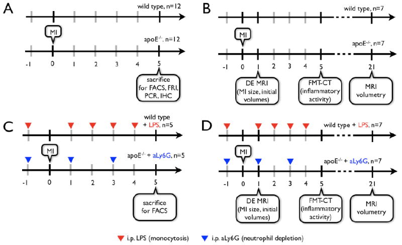
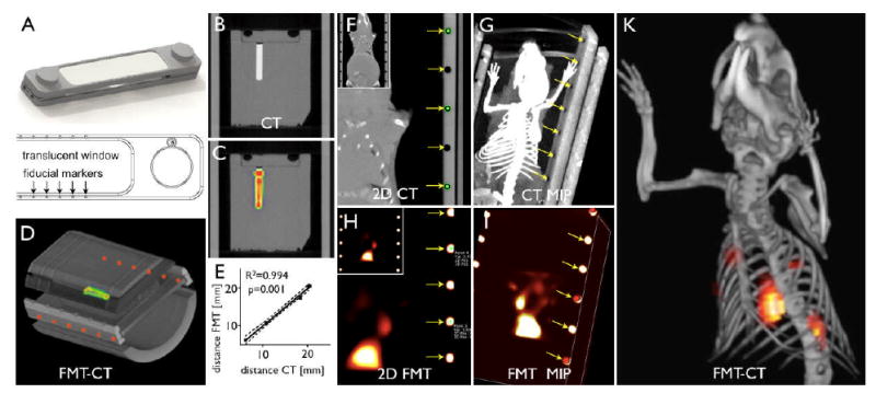
Methods: Ex vivo analysis of infarcts included flow cytometric monocyte enumeration, immunoactive staining, and quantitative polymerase chain reaction. To relate inflammatory activity to left ventricular remodeling, we used a combination of noninvasive fluorescence molecular tomography (FMT-CT) and physiologic imaging (magnetic resonance imaging).
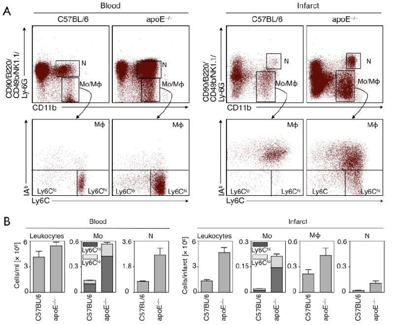
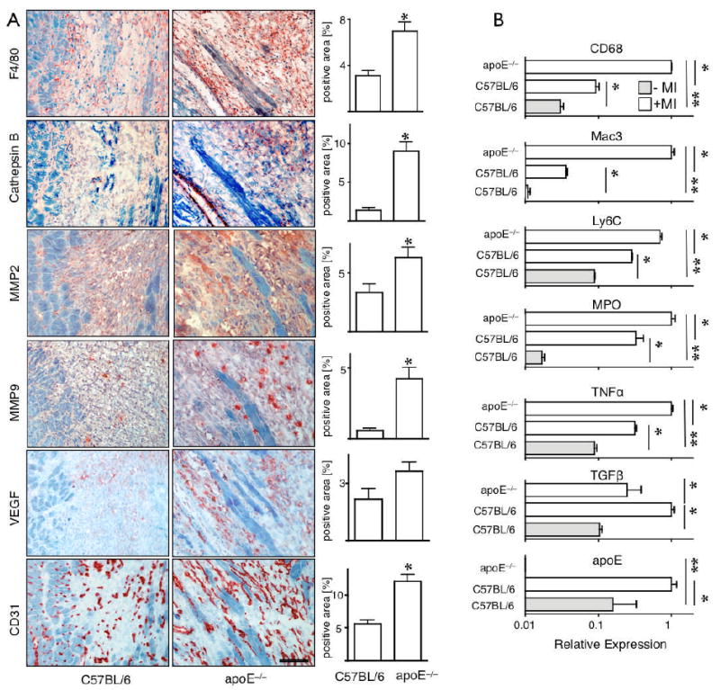
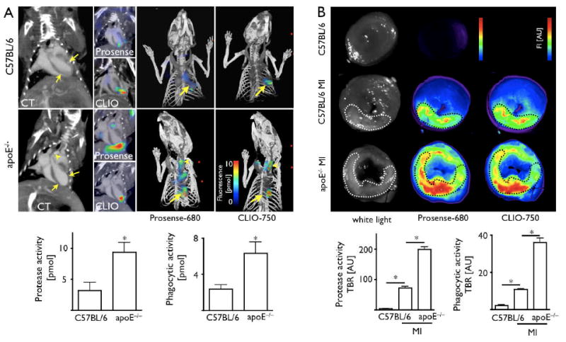
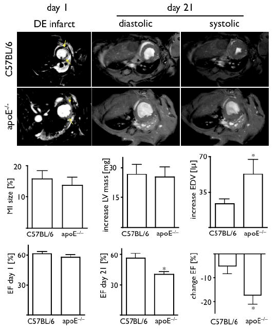
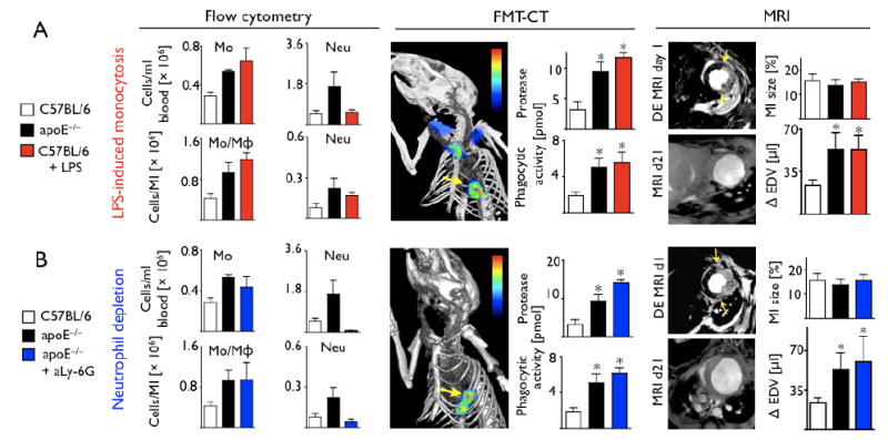
Results: Five-day-old infarcts showed >10x more Ly-6C(hi) monocytes in atherosclerotic (apoE(-/-)) mice compared with wild-type mice. The injured tissue in apoE(-/-) mice also showed a more pronounced inflammatory gene expression profile (e.g., increased tumor necrosis factor-alpha and myeloperoxidase and decreased transforming growth factor-beta) and a higher abundance of proteases, which are associated with the activity of Ly-6C(hi) monocytes. The FMT-CT on Day 5 after MI showed higher proteolysis and phagocytosis in infarcts of atherosclerotic mice. Serial magnetic resonance imaging showed accelerated deterioration of ejection fraction between Day 1 and Day 21 after MI in apoE(-/-). Finally, we could recapitulate these features in wild-type mice with artificially induced Ly-6C(hi) monocytosis.
Conclusions: Ly-6C(hi) monocytosis disturbs resolution of inflammation in murine infarcts and consequently enhances left ventricular remodeling. These findings position monocyte subsets as potential therapeutic targets to augment tissue repair after infarction and to prevent post-MI heart failure.
Copyright (c) 2010 American College of Cardiology Foundation. Published by Elsevier Inc. All rights reserved.
Conflict of interest statement
Figures
Comment in
-
When monocytes come (too) close to our hearts.J Am Coll Cardiol. 2010 Apr 13;55(15):1639-41. doi: 10.1016/j.jacc.2009.11.068. J Am Coll Cardiol. 2010. PMID: 20378084 No abstract available.
References
-
- Frangogiannis NG, Entman ML. Targeting the chemokines in myocardial inflammation. Circulation. 2004;110(11):1341–1342. - PubMed
-
- Blankesteijn WM, Creemers E, Lutgens E, Cleutjens JP, Daemen MJ, Smits JF. Dynamics of cardiac wound healing following myocardial infarction: observations in genetically altered mice. Acta Physiol Scand. 2001;173(1):75–82. - PubMed
-
- Cleutjens JP, Blankesteijn WM, Daemen MJ, Smits JF. The infarcted myocardium: simply dead tissue, or a lively target for therapeutic interventions. Cardiovasc Res. 1999;44(2):232–241. - PubMed
-
- Ertl G, Frantz S. Healing after myocardial infarction. Cardiovasc Res. 2005;66(1):22–32. - PubMed
-
- Sutton MG, Sharpe N. Left ventricular remodeling after myocardial infarction: pathophysiology and therapy. Circulation. 2000;101(25):2981–2988. - PubMed
Publication types
MeSH terms
Substances
Grants and funding
- R01 HL095612/HL/NHLBI NIH HHS/United States
- R24 CA092782/CA/NCI NIH HHS/United States
- U01 HL080731/HL/NHLBI NIH HHS/United States
- R01 AI084880/AI/NIAID NIH HHS/United States
- T32 CA079443/CA/NCI NIH HHS/United States
- R01HL096576/HL/NHLBI NIH HHS/United States
- P50 CA086355/CA/NCI NIH HHS/United States
- T32-CA79443/CA/NCI NIH HHS/United States
- R00 HL094533/HL/NHLBI NIH HHS/United States
- P50-CA86355/CA/NCI NIH HHS/United States
- R01 HL095629/HL/NHLBI NIH HHS/United States
- UO1-HL-080731/HL/NHLBI NIH HHS/United States
- R01-EB006432/EB/NIBIB NIH HHS/United States
- R01 EB006432/EB/NIBIB NIH HHS/United States
- R01 HL096576/HL/NHLBI NIH HHS/United States
- R01 HL093038/HL/NHLBI NIH HHS/United States
- R24-CA92782/CA/NCI NIH HHS/United States
LinkOut - more resources
Full Text Sources
Other Literature Sources
Medical
Research Materials
Miscellaneous
