. 2010 Jun 15;67(12):1128-36.
doi: 10.1016/j.biopsych.2010.02.008. Epub 2010 Apr 8.

Chronic stress causes amygdala hyperexcitability in rodents

Affiliations

Chronic stress causes amygdala hyperexcitability in rodents

J Amiel Rosenkranz et al. Biol Psychiatry. .

Abstract

Background: Chronic stress is a major health concern, often leading to depression, anxiety, or when severe enough, posttraumatic stress disorder. While many studies demonstrate that the amygdala is hyperresponsive in patients with these disorders, the cellular neurophysiological effects of chronic stress on the systems that underlie psychiatric disorders, such as the amygdala, are relatively unknown.

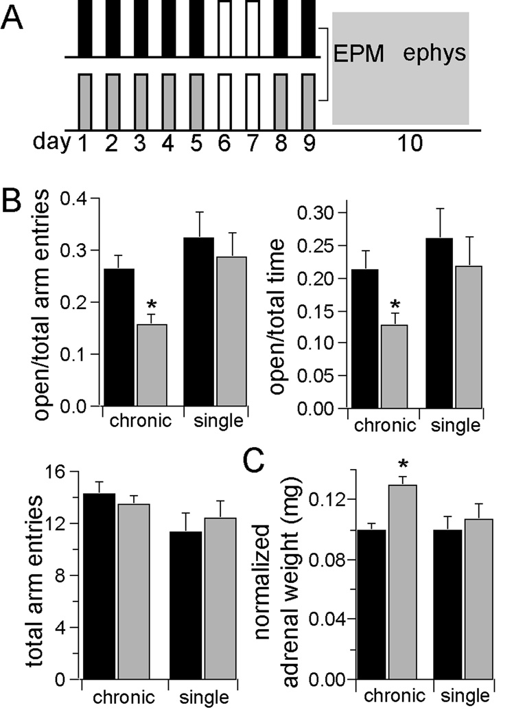
Methods: In this study, we examined the effects of chronic stress on the activity and excitability of amygdala neurons in vivo in rats. We used in vivo intracellular recordings from single neurons of the lateral amygdala (LAT) to measure neuronal properties and determine the cellular mechanism for the effects of chronic stress on LAT neurons.

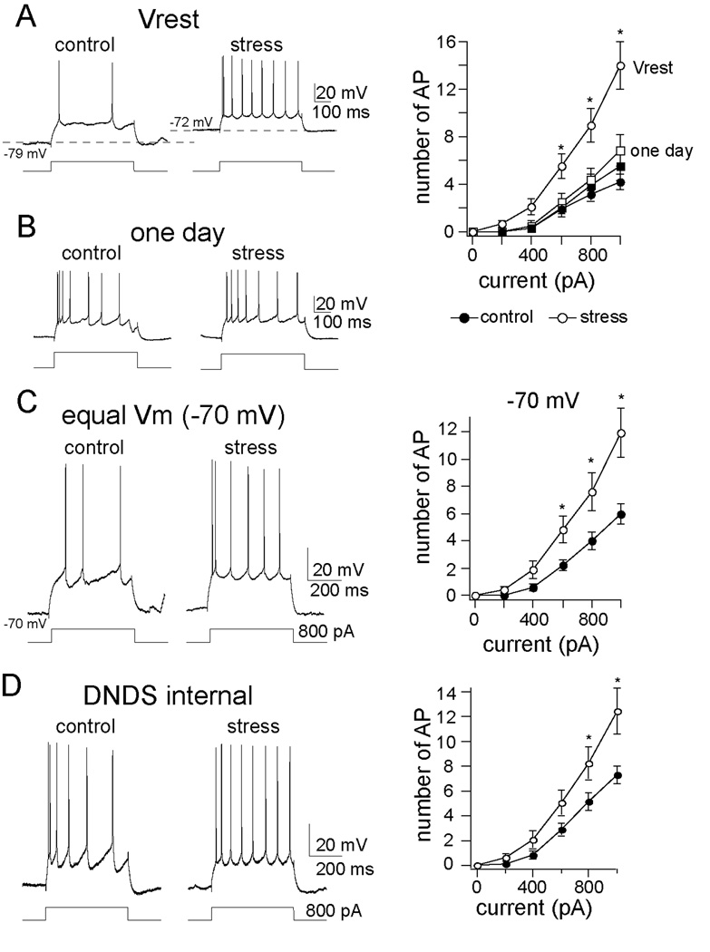
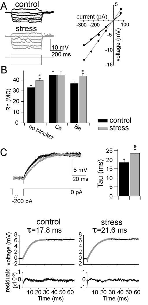
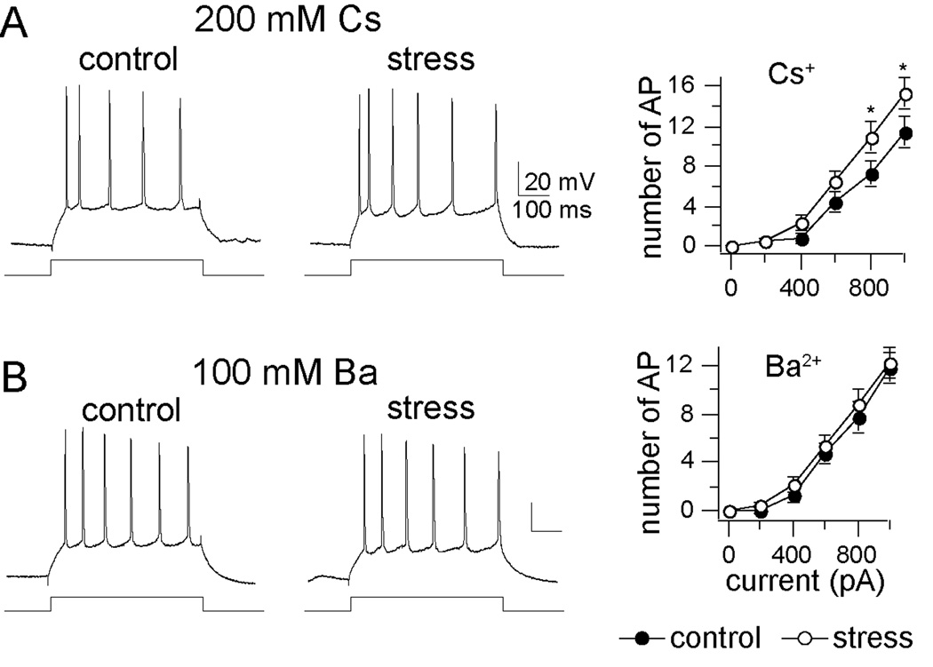
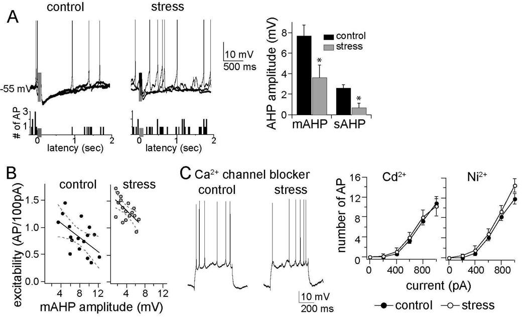
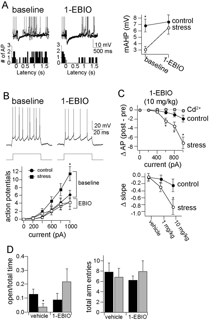
Results: We found a mechanism for the effects of chronic stress on amygdala activity, specifically that chronic stress increased excitability of LAT pyramidal neurons recorded in vivo. This hyperexcitability was caused by a reduction of a regulatory influence during action potential firing, facilitating LAT neuronal activity. The effects of stress on excitability were occluded by agents that block calcium-activated potassium channels and reversed by pharmacological enhancement of calcium-activated potassium channels.

Conclusions: These data demonstrate a specific channelopathy that occurs in the amygdala after chronic stress. This enhanced excitability of amygdala neurons after chronic stress may explain the observed hyperresponsiveness of the amygdala in patients with posttraumatic stress disorder and may facilitate the emergence of depression or anxiety in other patients.


Conflict of interest statement

The authors report no biomedical financial interests or potential conflicts of interest.

Figures

Figure 1
Figure 1. Repeated restraint is an effective chronic stressor
A) Rats were placed in a restraint chamber for 20 minutes, on 7 days over a 9 day period (gray). Control rats (black) experienced a similar degree of daily handling, but were not restrained. Following the last day of restraint, all rats were tested in the elevated plus maze. Some rats were then prepared for electrophysiology experiments. Remaining rats went through a fear conditioning and testing procedure. B) Repeated restraint stress decreased open arm entries (percent of open arm entries control 25.4 ± 2.7%, n=36, stress 15.6 ± 2.1%, n=34, p=0.0006, two-tailed t-test, t=3.58) and the time spent in open arms (percent of time in open arm, control 21.3 ± 2.9%, n=36, stress 12.8 ± 1.8%, n=34, p=0.016, two-tailed unpaired t-test, t=2.49), indicative of increased anxiety-like state. There was no significant change in the total number of arm entries (control 14.3 ± 0.9 arm entries, stress 13.5 ± 0.7 arm entries, p=0.485, two-tailed t-test, t=0.702). Single restraint did not significantly impact EPM exploration. C) Repeated restraint stress increased the weight of adrenal glands, a prototypical measure of the effectiveness of a stressor measured as raw weight (control 22.7 ± 0.7, n=12, stress 28.1 ± 1.2 mg, n=12, p=0.013, two-tailed unpaired t-test, t=2.92), or normalized to body weight (control 0.10 ± 0.005 mg/ kg, stress 0.13 ± 0.006 mg/ kg, p=0.002, two-tailed unpaired t-test, t=3.94). Single restraint did not significantly increase adrenal gland weight (normalized to body weight, control 0.10 ± 0.009, n=6, stress 0.11 ± 0.01, n=7, p=0.57, two-tailed unpaired t-test, t=0.61). * indicates significance at p<0.05.
Figure 2
Figure 2. Chronic stress increased the excitability of LAT neurons
A) Repeated restraint stress increased excitability measured from the neuronal resting membrane potential (Vrest; mixed design repeated measures ANOVA of each stimulation intensity, main effect of stimulation intensity F(5,264) = 31.67, p<0.001; main effect of stress F(1, 264) = 47.12, p<0.001; interaction F(5, 264) = 8.19, p<0.001). Shown here are voltage traces at the resting membrane potential of a neuron from the chronic handling control (left, Vrest = −79 m V; black circle in plot) and the chronic stress (right, Vrest = −72 m V, white circle in plot), in response to the same amplitude of current injection. B) The effect of chronic stress on excitability was only observed after repeated restraint, and not after a single restraint session (mixed design repeated measures ANOVA of each stimulation intensity, main effect of stress F(1, 72) = 0.79, p=0.376; main effect of stimulation intensity F(5,72) = 24.02, p<0.001; interaction F(5,72) = 0.20, p=0.96). This is measured as the response of these neurons to a depolarizing steps (squares in plot, one day control, black; one day stress, white). C) Repeated restraint stress caused an increase of LAT neuronal excitability when the membrane potential was held near −70 m V (mixed design repeated measures ANOVA of each stimulation intensity, main effect of stress F(1,264) = 22.19, p<0.001; main effect of stimulation intensity F(5,264) = 34.55, p<0.001; interaction F(5,264) = 3.39, p=0.005, * indicates p<0.05 between control and stress group in post-hoc unpaired t-tests with Bonferroni corrections). D) The effects of chronic stress on excitability were still observed when DNDS was included in the recording pipette (mixed design repeated measures ANOVA, main effect of stress F(1, 126) = 18.93, p<0.001, main effect of stimulation intensity F(5,126) = 47.59, p<0.001; control 0.88 ± 0.09 AP/ 100 pA, n=12; stress 1.67 ± 0.08 AP/ 100 pA, n=11, p<0.001, two-tailed unpaired t-test, t=6.51), demonstrating that the effects of chronic stress on excitability were not caused by a reduction of GABAergic inhibition.
Figure 3
Figure 3. Chronic stress altered membrane properties of LAT neurons
A) Repeated restraint (gray) caused an increase in the responsiveness to input, or input resistance (Rn), of LAT neurons (measured from −70 m V as the slope of the I–V relationship, control 33.3 ± 2.0 MOhms, n=21, stress 39.9 ± 2.1 MOhms, n=25, p=0.028, two-tailed unpaired t-test, t=2.275). B) Intracellular Cs+, but not Ba2+, blocked the effects of chronic stress on the Rn (Cs+ control 44.6 ± 3.3 MOhms, n=10, stress 45.2 ± 3.3 MOhms, n=10, p=0.899, two-tailed unpaired t-test, t=0.129; Ba2+ control 37.1 ± 2.2 MOhms, n=12, stress 43.9 ± 2.3 MOhms, n=11, p=0.041, two-tailed unpaired t-test, t=2.169). C) Chronic stress caused a lengthening of the membrane time constant (τ; control 18.1 ± 1.9 ms, stress 23.6 ± 1.9 ms, p=0.047, two-tailed unpaired t-test, t=1.80), as seen by the overlay of three decaying voltage responses after current injection in an example of a LAT neuron from control (black) and stress (grey) groups, and the time constant of the double exponential fit to this decay. * indicates significance at p<0.05.
Figure 4
Figure 4. Effects of single-cell block of K+ channels on excitability after chronic stress
A) Cs+ (200 mM; blocker of a variety of K+ channels) did not closely mimic the effects of chronic stress on excitability (mixed design repeated measures ANOVA of each stimulation intensity, main effect of stress F(1,108) = 10.79, p=0.0014; main effect of stimulation intensity F(5,108) = 58.98, p<0.001; interaction F(5,108) = 1.52, p=0.19). B) Ba2+ (100 mM), another K+ channel blocker that also blocks KCa channels, negated the effects of chronic stress on excitability (mixed design repeated measures ANOVA of each stimulation intensity, main effect of stress F(1,126) = 1.36, p=0.247; main effect of stimulation intensity F(5,126) = 64.03, p<0.001; interaction F(5,126) = 0.103, p=0.991). * indicates p<0.05 between control and stress group in post-hoc unpaired t-tests with Bonferroni corrections.
Figure 5
Figure 5. The AHP is reduced by chronic stress and necessary for the effects of chronic stress
A) The amplitudes of both the medium- and slow AHP were reduced after chronic stress (sAHP control 2.6 ± 0.3 m V, n=17, stress 0.7 ± 0.4 m V, n=18, p=0.003, two-tailed unpaired t-test, t=3.26; mAHP control 7.7 ± 1.1 m V, n=21, stress 3.6 ± 1.2 m V, n=25, p=0.009, two-tailed unpaired t-test, t=2.72), as seen in the neuronal response to a burst of 5 action potentials evoked by 5 current pulses (presented at grey box in overlay of 3 consecutive voltage traces and firing rate histogram; action potentials are truncated during the burst for clarity, but see Figure S2 in Supplement 1 for details). This reduction of the AHP amplitude was associated with greater spontaneous firing near action potential threshold, demonstrated in a firing rate histogram of 10 consecutive sweeps. B) The amplitude of the mAHP was correlated with neuronal excitability (control, r=−0.56, r2=0.32; stress, r=−0.62, r2=0.39), demonstrating that it is a major factor in regulating LAT neuronal activity. C) Intracellular administration of Ca2+ channel blockers mimicked the effects of chronic stress on neuronal excitability: Cd2+ (mixed design repeated measures ANOVA of each stimulation intensity, main effect of stress F(1,66) = 0.153, p=0.697; main effect of stimulation intensity F(5,66) = 40.72, p<0.001; interaction F(5,66) = 0.124, p=0.987) and Ni2+ (mixed design repeated measures ANOVA of each stimulation intensity, main effect of stress F(1,72) = 3.77, p=0.056; main effect of stimulation intensity F(5,72) = 86.33, p<0.001; interaction F(5,72) = 0.711, p=0.617). Displayed 28 in this panel are traces with intracellular Cd 2+. * indicates p<0.05 between control and stress group in post-hoc unpaired t-tests with Bonferroni corrections.
Figure 6
Figure 6. Pharmacological enhancement of the AHP reversed the effects of chronic stress on excitability and EPM
A) 1-EBIO is an activator of KCa channels. Administration of 1-EBIO increased the amplitude of the AHP, and augmented the impact of the AHP on spontaneous action potential firing, demonstrated in a firing rate histogram of 10 consecutive sweeps. Displayed here are overlays of three consecutive traces before and after 1-EBIO (10 mg/ kg, i.p.). 1-EBIO brought the amplitude of the mAHP to near-control levels. B) 1-EBIO causes a reduction in the excitability of LAT neurons. C) The effects of 1-EBIO (10 mg/ kg) were greater in chronic stress animals, and returned neuronal excitability to close to control, non-stress levels. These effects were also dose-dependent, and blocked by intracellular application of Cd2+ (0.5 mM; mixed design repeated measure ANOVA, main effect of 1-EBIO F(1,48) = 0.047, p=0.829, main effect of stimulation intensity F(5,48) = 24.46, p<0.001), indicating that its effects on LAT neuronal excitability are caused by direct actions on LAT neurons. D) 1-EBIO (10 mg/ kg) reversed the effects of chronic stress on exploration in the EPM, as demonstrated by increased time spent in the open arms (percent time in open arms, Kruskal-Wallis = 8.22, p=0.042, n=8/ group; vehicle control 12.7 ± 3.7%, vehicle chronic stress 3.8 ± 2.3%, Mann-Whitney U = 10.0, p=0.023; 1-EBIO control 8.6 ± 4.6%, 1-EBIO chronic stress 21.8 ± 8.9%, Mann-Whitney U = 22.0, p=0.33). There was no significant effect on the total number of arm entries (Kruskal-Wallis = 0.198, p = 0.978, n=8/ group; control vehicle 7.8 ± 1.7 entries, stress vehicle 6.8 ± 1.7 entries; control 1-EBIO 6.1 ± 0.8 entries, stress 1-EBIO 7.9 ± 2.4 entries).

Comment in

Similar articles

Cited by

References

    1. Chida Y, Hamer M. Chronic psychosocial factors and acute physiological responses to laboratory-induced stress in healthy populations: a quantitative review of 30 years of investigations. Psychol Bull. 2008;134:829–885. - PubMed
    1. Pohl J, Olmstead MC, Wynne-Edwards KE, Harkness K, Menard JL. Repeated exposure to stress across the childhood-adolescent period alters rats' anxiety- and depression-like behaviors in adulthood: The importance of stressor type and gender. Behav Neurosci. 2007;121:462–474. - PubMed
    1. Wood GE, Norris EH, Waters E, Stoldt JT, McEwen BS. Chronic immobilization stress alters aspects of emotionality and associative learning in the rat. Behav Neurosci. 2008;122:282–292. - PubMed
    1. Heim C, Nemeroff CB. The role of childhood trauma in the neurobiology of mood and anxiety disorders: preclinical and clinical studies. Biol Psychiatry. 2001;49:1023–1039. - PubMed
    1. Lupien SJ, McEwen BS, Gunnar MR, Heim C. Effects of stress throughout the lifespan on the brain, behaviour and cognition. Nat Rev Neurosci. 2009;10:434–445. - PubMed

Publication types

MeSH terms

Substances