Review
. 2010 Dec;94(7):2507-13.
doi: 10.1016/j.fertnstert.2010.02.037. Epub 2010 Apr 7.

Oocyte maturation failure: a syndrome of bad eggs

Affiliations
Review

Oocyte maturation failure: a syndrome of bad eggs

Stephanie Beall et al. Fertil Steril. 2010 Dec.

Abstract

To show that disruption of meiotic competence results in cell cycle arrest, and the production of immature oocytes that are not capable of fertilization. Through an extensive review of animal studies and clinical case reports, we define the syndrome of oocyte maturation failure as a distinct oocyte disorder, present a classification system based on clinical parameters, and discuss the potential molecular origins for the disease.


Figures

FIGURE 1
FIGURE 1
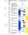
Key molecular events during meiotic progression. Oocyte meiosis initiates during in utero development when the female germ cell progresses through prophase I and arrests at the diplotene stage before birth. During this time, the chromosomes condense, cohesion between sister chromatids is established and synaptonemal complexes form between homologous chromosomes. The cohesin proteins form the molecular glue between sister chromatids and proteins involved in double-strand break repair in addition to meiosis, specific SCP1, 2, and 3 are essential for recombination and synaptonemal complex formation. During the reproductive phase of life, reinitiation of meiosis occurs in response to hormonal stimulation just before ovulation. The teeter-totter between oocyte arrest and progression depends on alternating levels of the key mediators: MPF and APC/C. This is regulated by multiple signaling cascades as illustrated above. LH, through a mechanism not yet elucidated, stimulates PDE3 resuting in decresed cAMP levels within the oocyte. Simultaneously, cdc25 is activated. The combination of these two events results in activation of MPF, which drives meiotic progression. Exit out of prophase I and entry into M phase of meiosis I is hallmarked by the GVBD and the formation of the meiotic spindle. Following formation of the spindle apparatus homologous chromosome pairs align along the meiotic equatorial plate. When the chromosomes are all properly aligned, the SAC signal is extinguished, resulting in APC/C activation. The APC/C is a ubiquitin ligase that targets securing for degradation allowing the release of the active form of separase, which cleaves Rec 8, therefore releasing the cohesin glue along chromosome arms resulting in separation homologous chromosomes. Sister chromatids do not separate at this point in meiosis because Rec8 at the centromere is protected by shugosin during metaphase I. Following the separation of the sister chromosomes, Emi2, through its inhibition of APC/C maintains MPF function, therefore allowing progression of meiosis II through to metaphase II arrest. The oocyte remains arrested at this stage until it is fertilized. Upon sperm penetration, a rise in intracellular calcium concentration leads to CamKII activation and subsequent inhibition of Emi2. This chain of events leads to the activation of APC/C. In a process similar to the metaphase I-anaphase I transition, APC/C activation results in destruction of MPF and the release of separase. Centromeric Rad8 is no longer protected by shugosin, therefore allowing cleavage of cohesin at the centromere and sister chromatid separation. Meiosis is complete following reformation of the nuclear envelope, uncoiling of chromosomes and second polar body formation. At this point the gamete transitions from meiotic cell division to mitotic, thus permitting embryonic development.

Similar articles

Cited by

References

    1. Muasher SJ, Abdallah RT, Hubayter ZR. Optimal stimulation protocols for in vitro fertilization. Fertil Steril. 2006;86:267–73. - PubMed
    1. Ebner T, Moser M, Sommergruber M, Tews G. Selection based on morphological assessment of oocytes and embryos at different stages of preimplantation development: a review. Hum Reprod Update. 2003;9:251–62. - PubMed
    1. Bar-Ami S, Zlotkin E, Brandes JM, Itskovitz-Eldor J. Failure of meiotic competence in human oocytes. Biol Reprod. 1994;50:1100–7. - PubMed
    1. Avrech OM, Goldman GA, Rufas O, Stein A, Amit S, Yoles I, et al. Treatment variables in relation to oocyte maturation: lessons from a clinical micromanipulation-assisted in vitro fertilization program. Assist Reprod Genet. 1997;14:337–42. - PMC - PubMed
    1. Jamnongjit MHS. Oocyte maturation: the coming of ageofa germ cell. Semin Reprod Med. 2005;23:234–41. - PMC - PubMed