. 2010 Jun 11;285(24):18640-9.
doi: 10.1074/jbc.M109.085746. Epub 2010 Apr 8.

The integrin co-activator Kindlin-3 is expressed and functional in a non-hematopoietic cell, the endothelial cell

Affiliations

The integrin co-activator Kindlin-3 is expressed and functional in a non-hematopoietic cell, the endothelial cell

Katarzyna Bialkowska et al. J Biol Chem. .

Abstract

Integrin activation is crucial for numerous cellular responses, including cell adhesion, migration, and survival. Recent studies in mice have specifically emphasized the vital role of kindlin-3 in integrin activation. Kindlin-3 deficiency in humans also has now been documented and includes symptoms of bleeding, frequent infections, and osteopetrosis, which are consequences of an inability to activate beta1, beta2, and beta3 integrins. To date, kindlin-3 was thought to be restricted to hematopoietic cells. In this article, we demonstrate that kindlin-3 is present in human endothelial cells derived from various anatomical origins. The mRNA and protein for KINDLIN-3 was detected in endothelial cells by reverse transcription-PCR and Western blots. When subjected to sequencing by mass spectrometry, the protein was identified as authentic kindlin-3 and unequivocally distinguished from KINDLIN-1 and KINDLIN-2 or any other known protein. By quantitative real time PCR, the level of kindlin-3 in endothelial cells was 20-50% of that of kindlin-2. Using knockdown approaches, we show that kindlin-3 plays a role in integrin-mediated adhesion of endothelial cells. This function depends upon the integrin and substrate and is distinct from that of kindlin-2. Formation of tube-like structures in Matrigel also was impaired by kindlin-3 knockdown. Mechanistically, the distinct functions of the kindlins can be traced to differences in their subcellular localization in integrin-containing adhesion structures. Thus, the prevailing view that individual kindlins exert their functions in a cell type-specific manner must now be modified to consider distinct functions of the different family members within the same cell type.


Figures

FIGURE 1.
FIGURE 1.
Expression of KINDLIN-3 mRNA in endothelial cells. 200 ng of total RNA was subjected to RT-PCR, using KINDLIN-2- and KINDLIN-3-specific primers. GAPDH was used as a loading control.
FIGURE 2.
FIGURE 2.
Identification of kindlin-3 protein in endothelial cells by Western blotting and immunoprecipitation. A, Western blots of CHO cells expressing EGFP-tagged kindlin-1, kindlin-2, or kindlin-3 cell lysates were probed with kindlin-3 antibodies (upper panel) or EGFP monoclonal antibody (lower panel). The specificity of the anti-kindlin-3 anti-peptide antibody is demonstrated. B, Western blots of HUVECs, Meg-01, platelet, and lymphocyte cell lysates were probed with the anti-peptide kindlin-3 antibodies. Identical results were obtained in three independent experiments. C, Western blots of HUVEC and platelet lysates were developed with the anti-peptide kindlin-3 antibodies and GAPDH antibodies. D, Western blots of HUVEC, brain, heart, and liver extracts were probed with the anti-peptide kindlin-3 antibodies and GAPDH antibodies. E, Western blots of HUVECs and CHO cells expressing EGFP-kindlin-3 cell lysates were probed with anti-peptide kindlin-3 antibodies in the absence (left-hand panel) or presence (right-hand panel) of the immunizing peptide. F, HUVECs or Meg-01 cell lysates were immunoprecipitated with kindlin-3 antibodies. Preimmune serum was used as negative control. Immunoprecipitates were analyzed on Western blots with a second kindlin-3 antibody. Identical results were obtained in three independent experiments.
FIGURE 3.
FIGURE 3.
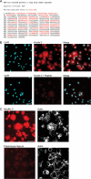
Detection of kindlin-3 in endothelial cells by mass spectrometry and immunofluorescence. A, endogenous kindlin-3 was immunoprecipitated from HUVEC with peptide antibodies. The 72-kDa band was excised from the gel and subjected to mass spectrometry sequencing. Peptide coverage is shown, matched peptides shown in red. Peptide coverage was 41%. B, HUVECs spread on fibrinogen for 30 min were fixed, permeabilized, and stained with peptide antibodies to detect kindlin-3, followed by Alexa 568 anti-rabbit IgG and DAPI to visualize all the cells (upper panels). The specificity of the anti-kindlin-3 peptide antibodies was examined by inclusion of immunizing peptide in the primary antibody labeling step (lower panels). C, HUVECs spread on vitronectin for 1 h were fixed, permeabilized, and stained with peptide antibodies to detect kindlin-3, followed by Alexa 568 anti-rabbit IgG and phalloidin-Alexa 633 to visualize all the cells (upper panels). Preimmune serum from the same rabbit was used for staining to establish background staining (lower panels). Bars = 40 μm.
FIGURE 4.
FIGURE 4.
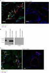
Kindlin-3 is present in endothelial cells in vivo. A, frozen tissue sections of human prostate cancer were stained for kindlin-3 (green) and CD31 (red) and mounted in DAPI containing medium. Endothelial cells positive for CD31 and kindlin-3 are marked with arrows. The kindlin-3 immunizing peptide used to raise the antibodies was included as specificity controls. B, Western blots of mouse and human platelet and human lymphocyte cell lysates were probed with the anti-peptide kindlin-3 antibodies in the absence (left-hand panel) or presence (right-hand panel) of the immunizing peptide. C, frozen tissue sections of mouse skeletal muscle were stained for kindlin-3 (green) and CD31 (red) and mounted in DAPI containing medium. Endothelial cells positive for CD31 and kindlin-3 are marked with arrows. The kindlin-3 immunizing peptide used to raise the antibodies was included as specificity controls. Bars = 20 μm.
FIGURE 5.
FIGURE 5.
Kindlin-3 distribution in HUVECs spread on integrin substrates. HUVECs spread on vitronectin (VN) and fibrinogen (FIB) (A) for 1 h or fibronectin (FN) and collagen (COL) (B) for 2 or 1 h, respectively, were fixed, permeabilized, and stained with antibodies to detect kindlin-2, kindlin-3, β3 integrin, and β1 integrin followed by Alexa 633 anti-mouse IgG, Alexa 568 anti-rabbit IgG, and Alexa 488 anti-rat IgG. Kindlin-3 colocalized with integrins at the sites of membrane extensions (large arrows) but not in the focal adhesions (small arrows). Kindlin-2 colocalized with integrins at sites of membrane extension and in focal adhesions (double arrows). C, merge of kindlin-2 and kindlin-3 staining from A is shown. Areas rich in kindlin-2 and devoid of kindlin-3 are marked with boxes. Bars = 20 μm. The images shown are representative of 12–15 independent scans for each integrin ligand.
FIGURE 6.
FIGURE 6.
Kindlin-3 knockdown inhibits integrin-mediated adhesion in HUVECs. A, siRNA suppression of kindlin-3 expression in HUVECs. Expression of kindlin-2 and kindlin-3 in non-transfected cells (NT), kindlin-2 (Kin2), kindlin-3 (Kin3), and non-targeting siRNA (C) transfectants were analyzed by Western blotting with kindlin-2, kindlin-3, and GAPDH antibodies. B and C, non-transfected or HUVECs transfected with kindlin-2 (Kin2), kindlin-3 (Kin3), or non-targeting siRNA (C) were used in adhesion assays. The cells were fixed, stained with DAPI and Alexa 568-phalloidin, and counted. The error bars are mean ± S.E. of three independent experiments.
FIGURE 7.
FIGURE 7.
Kindlin-3 knockdown inhibits formation of tube-like structures in vitro. Non-transfected, non-targeting siRNA-transfected or kindlin-3 siRNA-transfected HUVECs were seeded on growth factor-reduced Matrigel containing 10 μg/ml of fibronectin and 10 ng/ml of vascular endothelial growth factor. The figure shows representative fields of three separate experiments, each performed in triplicate. Bar = 200 μm.

References

    1. Qin J., Vinogradova O., Plow E. F. (2004) PLoS Biol. 2, 726–729 - PMC - PubMed
    1. Ma Y. Q., Qin J., Plow E. F. (2007) J. Thromb. Haemost. 5, 1345–1352 - PubMed
    1. Tu Y., Wu S., Shi X., Chen K., Wu C. (2003) Cell 113, 37–47 - PubMed
    1. Ussar S., Wang H. V., Linder S., Fässler R., Moser M. (2006) Exp. Cell Res. 312, 3142–3151 - PubMed
    1. Shi X., Ma Y. Q., Tu Y., Chen K., Wu S., Fukuda K., Qin J., Plow E. F., Wu C. (2007) J. Biol. Chem. 282, 20455–20466 - PubMed

Publication types

MeSH terms