. 2010 Jun 18;285(25):19502-9.
doi: 10.1074/jbc.M109.098186. Epub 2010 Apr 8.

Molecular mechanism that induces activation of Spätzle, the ligand for the Drosophila Toll receptor

Affiliations

Molecular mechanism that induces activation of Spätzle, the ligand for the Drosophila Toll receptor

Christopher J Arnot et al. J Biol Chem. .

Abstract

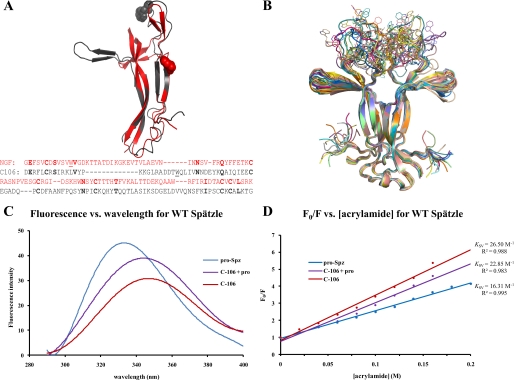
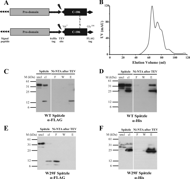
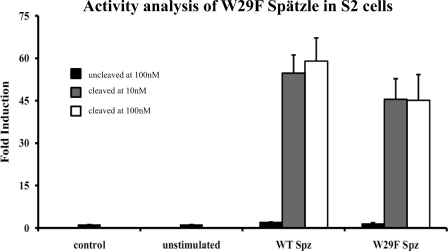
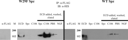
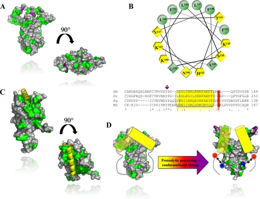
The Drosophila Toll receptor is activated by an endogenous cytokine ligand Spätzle. Active ligand is generated in response to positional cues in embryonic dorso-ventral patterning and microbial pathogens in the insect immune response. Spätzle is secreted as a pro-protein and is processed into an active form by the serine endoproteases Easter and Spätzle-processing enzyme during dorso-ventral patterning and infection, respectively. Here, we provide evidence for the molecular mechanism of this activation process. We show that the Spätzle prodomain masks a predominantly hydrophobic region of Spätzle and that proteolysis causes a conformational change that exposes determinants that are critical for binding to the Toll receptor. We also gather that a conserved sequence motif in the prodomain presents features of an amphipathic helix likely to bind a hydrophobic cleft in Spätzle thereby occluding the putative Toll binding region. This mechanism of activation has a striking similarity to that of coagulogen, a clotting factor of the horseshoe crab, an invertebrate that has changed little in 400 million years. Taken together, our findings demonstrate that an ancient passive defense system has been adapted during evolution and converted for use in a critical pathway of innate immune signaling and embryonic morphogenesis.


Figures

FIGURE 1.
FIGURE 1.
Proteolytic activation of Spätzle induces a conformational change. A, structural and sequence alignment of nerve growth factor (residues 131–236, gray) and Spätzle C-106 (arbitrary sequence numbers because of N-terminal splice variant) (see “Experimental Procedures”). Tryptophan residues are illustrated as spheres, and the structures have a C-α backbone root means square deviation of 2.72 Å. In the sequence alignment, identical residues are in bold, and the tryptophans are underlined. B, C-106 dimer structure (18). The tryptophan loop is shown in 20 possible conformations selected on the basis of energy minimization. C, fluorescence spectroscopy of Spätzle (see “Experimental Procedures”). Red shifting of the spectra shows that the tryptophan residue is increasingly exposed as the prodomain is cleaved but remains noncovalently attached (C-106 + pro) and then most exposed once the prodomain is completely removed (C-106). D, quenching of the fluorescence signal using acrylamide. This allows for the calculation of the Stern–Volmer constant (Ksv), which gives a quantitative indication of the level of exposure of the tryptophan residue; the higher Ksv values indicate increased exposure of the residue.
FIGURE 2.
FIGURE 2.
Properties of W29F Spätzle. A, schematic of the Spätzle expression construct with TEV cleavage site, FLAG, and His epitope tags is shown. B, elution profile of W29F Spätzle separated by gel filtration on Superdex S-200 is shown. C and D, wild-type (WT) C-106 elutes along with the His-tagged prodomain following cleavage by TEV protease and subsequent Ni-NTA purification showing noncovalent interaction. E and F, W29F mutation causes C-106 to elute in the flow-through following Ni-NTA purification, implying a decreased interaction between the prodomain and C-106. The prodomain and C-106 are detected with anti-His and anti FLAG antibodies, respectively.
FIGURE 3.
FIGURE 3.
W29F C-106 activates Toll signaling with characteristics similar to the wild-type protein. S2 cells expressing a drosomycin-luciferase reporter construct were cultured for 24 h in tissue culture medium supplemented with 10 nm or 100 nm Spätzle. After 24 h, cells were lysed, and luciferase activity was measured in triplicate (see “Experimental Procedures”). Data represent mean ± S.D. (error bars) and are shown as fold induction.
FIGURE 4.
FIGURE 4.
Uncleaved W29F Spätzle pro-protein binds to the Toll ectodomain. Immunoprecipitation experiments were conducted by binding either uncleaved (Spz) or TEV protease-cleaved (C106) to the immunoprecipitation matrix using anti-FLAG antibody, then incubating with His-tagged Toll extracellular domain to test for interaction. The reducing gels were then probed with anti-His antibody. Left, W29F mutation in C-106 allows for Toll extracellular domain (ECD) binding of both uncleaved and cleaved Spätzle. Right, as expected, uncleaved Trp29 Spätzle does not bind to Toll, whereas wild-type (WT) C-106 does.
FIGURE 5.
FIGURE 5.
An intact Trp loop is not required for Toll binding. A, Spätzle pro-protein was overdigested with trypsin, and the mixture was separated by ion-exchange chromatography (Mono Q). The four peaks were analyzed by reducing and nonreducing SDS-PAGE. Full-length C-106 is marked with a line (24 kDa nonreduced, 12 kDa reduced), whereas C-106 cleaved at Arg24 is indicated by an asterisk (8.9-kDa reduced fragment) and a triangle (3.1-kDa reduced fragment). B, Toll ectodomain was mixed with C-106 cleaved at Arg24. The Toll and cleaved C-106 co-elute at 11.1 ml as an apparent dimer on a Superdex S-200 (GE Healthcare) (see also Ref. 8).
FIGURE 6.
FIGURE 6.
Conserved regulatory a-helix in Spätzle and horseshoe crab coagulogen. A, the molecular surface of C-106 with hydrophobic patches. Hydrophobic residues (alanine, glycine, valine, isoleucine, leucine, phenylalanine, methionine) are colored in green. The tryptophan residue Trp29 is shown in purple. B, lower, sequence alignment of the Spätzle prodomains from D. melanogaster (Dm), D. virilis (Dv), A. gambiae (Ag), and M. sexta (Ms). The position of the predicted α-helix is indicated by a yellow cylinder. A purple arrow indicates the position of the two Spätzle null mutations (Y134N, P135L). B, upper, helical-wheel projection of residues 132–157. In yellow hexagons are positively charged residues. Yellow diamonds are hydrophilic, and green positions are hydrophobic residues. C, crystal structure of coagulogen (PDB code 1AOC) with the same color scheme as in A. The regulatory helix is shown in yellow. D, schematic mechanism for Spätzle activation. Prior to activation, the conserved prodomain helix masks Trp29 and the Toll binding sites of Spätzle. Proteolysis induces a conformational change that partially exposes the Toll-binding determinants. Receptor binding displaces the prodomain otherwise tightly bound to C-106 via a charge clamp between the newly formed N terminus (in blue) and the conserved aspartate (in red) C terminus of the prodomain α-helix.

Similar articles

Cited by

References

    1. Belvin M. P., Anderson K. V. (1996) Annu. Rev. Cell Dev. Biol. 12, 393–416 - PubMed
    1. Hoffmann J. A. (2003) Nature 426, 33–38 - PubMed
    1. Schneider D. S., Jin Y., Morisato D., Anderson K. V. (1994) Development 120, 1243–1250 - PubMed
    1. Weber A. N., Tauszig-Delamasure S., Hoffmann J. A., Lelièvre E., Gascan H., Ray K. P., Morse M. A., Imler J. L., Gay N. J. (2003) Nat. Immunol. 4, 794–800 - PubMed
    1. Mizuguchi K., Parker J. S., Blundell T. L., Gay N. J. (1998) Trends Biochem. Sci. 23, 239–242 - PubMed

LinkOut - more resources