. 2010 Aug;38(14):4620-34.
doi: 10.1093/nar/gkq228. Epub 2010 Apr 8.

Recruitment of MBD1 to target genes requires sequence-specific interaction of the MBD domain with methylated DNA

Affiliations

Recruitment of MBD1 to target genes requires sequence-specific interaction of the MBD domain with methylated DNA

Thomas Clouaire et al. Nucleic Acids Res. 2010 Aug.

Abstract

MBD1, a member of the methyl-CpG-binding domain family of proteins, has been reported to repress transcription of methylated and unmethylated promoters. As some MBD1 isoforms contain two DNA-binding domains-an MBD, which recognizes methylated DNA; and a CXXC3 zinc finger, which binds unmethylated CpG-it is unclear whether these two domains function independently of each other or if they cooperate in facilitating recruitment of MBD1 to particular genomic loci. In this report we investigate DNA-binding specificity of MBD and CXXC3 domains in vitro and in vivo. We find that the methyl-CpG-binding domain of MBD1 binds more efficiently to methylated DNA within a specific sequence context. We identify genes that are targeted by MBD1 in human cells and demonstrate that a functional MBD domain is necessary and sufficient for recruitment of MBD1 to specific sites at these loci, while DNA binding by the CXXC3 motif is largely dispensable. In summary, the binding preferences of MBD1, although dependent upon the presence of methylated DNA, are clearly distinct from those of other methyl-CpG-binding proteins, MBD2 and MeCP2.


Figures
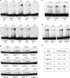
Figure 1.
Figure 1.
Binding specificity of MBD and CXXC3 domains of MBD1 in vitro. (A) EMSA combined with base substitution scanning mutagenesis of nucleotides adjacent to methylated CpG detect preferential binding of MBD domain to probes with T in position −1 and C in position +1. Lanes labelled with ‘−’ contain no protein. The triangle indicates increasing concentrations of MBD (75 and 200 nM). All probes used in these experiments happen to contain an A at position +2 from the CG. Therefore MBD1 binds seemingly identical probes such as TMCGG and CMCGA or GMCGG and CMCGC with different efficiency. (B) The CXXC3 domain binds with similar efficiency to all unmethylated probes containing a CG pair independently of the sequence context, but does not bind to methylated probes. Lanes labelled with ‘−’ contain no protein. The triangle indicates increasing amounts of CXXC3 (50–200 nM). (C) The MBD domain of MBD1 (M1) but not the MBD domain of MeCP2 (Me2) efficiently binds TGMCGCA sequence enriched in methyl-SELEX experiment and probe containing TMCGC from base substitution scanning mutagenesis with A at position +2. 200 nM of M1 and Me2 were used in these EMSA experiments. (D) Replacement of A to C at position +2 in TMCGCA sequence reduces the affinity of MBD1 MBD domain to methylated probe. Replacement of T at position −2 with G does not affect the efficiency of binding. (E) Relative KD of MBD1 MBD binding to probes containing the optimal (TMCGCA) or suboptimal methylated binding sites. Note that the MBD domain binds TMCGCA probe 3–5-fold more efficiently than any other methylated sequence.
Figure 2.
Figure 2.
Mutations in MBD and CXXC3 domains disrupt DNA binding. (A) Schematic representation of PCM1 variant of MBD1. The MBD domain, CXXC zinc fingers and transcriptional repression domain (TRD) are indicated. The solution structure of the MBD (43) and CXXC3 domains (32) is shown as well as the position of amino acids, which we have mutated, contributing to DNA binding (red) or coordination of zinc (blue). (B) Replacement of arginine (R) 22 to alanine (A) disrupts binding of MBD to methylated DNA. 50, 100, 200 and 400 nM of MBD were used in EMSA experiments with methylated probes and 100, 200 and 400 nM for EMSA with unmethylated probes. (C and D) Mutations of zinc coordinating cysteines (C289 and C292) to alanine and lysines (K310, K312 and K319) to alanine within the DNA binding part of CXXC3 disrupt binding of CXXC3 to DNA. The triangles represent 5-fold increments in CXXC3 concentration in EMSA experiments. In control lanes indicated with ‘−’ no protein was used. In lanes marked with M+ methylated DNA probes were used in EMSA experiments.
Figure 3.
Figure 3.
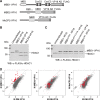
MBD1-VP16 fusion activates specific genes in HeLa cells. (A) The fusion proteins containing DNA binding domains of MBD1, MBD2 and MeCP2 and transactivation domain of herpes virus (VP16 AD). All proteins have a C-terminal FLAG peptide. (GR)11 is a region of MBD2 rich in glycine and arginine. (B) Expression of MBD-VP16 fusion proteins in HeLa cells. Histone deacetylase (HDAC1) detected simultaneously with the fusion proteins serves as a loading control. (C) Expression of MBD1-VP16 and MBD1-VP16 proteins carrying point mutations in either the MBD (R22A) or CXXC3 (K310,319A and C289,292A) domains. MBD1DM-VP16 combines R22A with C289,292A mutations. HDAC1 serves as a loading control. (D) Log 2 plots show the effect of MBD-VP16 fusion proteins on transcription in HeLa cells. In all graphs the x-axis represents M = log 2 MBD-VP16/HeLa and the y-axis, the control, M = log 2 MBD1DM-VP16/HeLa. Transcripts up-regulated by MBD1-VP16 3-fold or more (ΔM ≥ 1.5) are shown in red. Note that transcripts induced by MBD1-VP16 are not up-regulated after expression of either MBD2-VP16 or MeCP2-VP16.
Figure 4.
Figure 4.
Validation of microarray data and characterization of transactivation potential of mutant MBD1-VP16 proteins. (A and B) Semiquantitative and quantitative RT-PCR experiments demonstrate that transcripts induced by expression of MBD1-VP16 in HeLa by ∼10-fold are not up-regulated in cells expressing either MBD2-VP16 or MeCP2-VP16. Actin served as ubiquitously expressed control. The graphs in (B) represent triplicate RT-qPCRs performed on two independent transfection experiments for each fusion protein. (C and D) Semi-quantitative and quantitative RT-PCRs show that MBD1-VP16 carrying R22A a mutation in the MBD domain (R22A and DM) is unable to activate target genes compared to MBD1-VP16 carrying K310,319A mutations in CXXC3. Cysteine mutations (C289A,292A) also abolished transactivation by MBD1-VP16, presumably by affecting protein folding. Actin was used as a control. RT-qPCR experiments were performed in triplicates on two independent transfections for each protein. The expression of MBD1-, MBD2- and MeCP2-VP16 fusion proteins as well as expression of mutant forms of MBD1-VP16 is shown in Figure 3B and C.
Figure 5.
Figure 5.
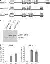
Lack of CXXC3 domain does not affect transactivation by MBD1-VP16 and binding of MBD1 to DNA in living cells. (A) Schematic representation of MBD1 isoforms Variant1 (MBD1Var1), Variant3 (MBD1Var3) and PCM1 (MBD1PCM1). Note that MBD1Var1 carries three CXXC motifs, while MBD1Var3 lacks DNA binding CXXC3 and MBD1PCM1 lacks CXXC2. (B) Expression of MBD1PCM1-VP16 and MBD1Var3-VP16 in HeLa cells. HDAC1 serves as a loading control. (C) RT-qPCR experiments demonstrate that MBD1PCM1-VP16 (M1) and MBD1Var3-VP16 (M1Var3) do not differ significantly in their ability to induce HBA and RND transcription in HeLa.
Figure 6.
Figure 6.
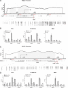
Chromatin immunoprecipitation detects MBD1 binding near RND2 and NGFR promoters. (A) Schematic drawing of RND2 promoter with transcription start site (TSS) indicated by arrow. The GC content, position of CpGs and regions analysed by ChIP in (B) are shown. The promoter sequences from −643 to +825 were analysed for DNA methylation by bisulphite sequencing. The results are displayed below the line corresponding to the sequenced region. Filled circles represent methylated CpGs. The red bar in the CpG plot at +801 and the red dot under the bisulphite sequencing plot indicate a high affinity MBD1 binding site (TCGCA). (B) qPCRs on chromatin immunoprecipitations (ChIP) with antibodies against VP-16AD and MBD1 detect binding of MBD1 and MBD1-VP16, but not DM-VP16, at high affinity MBD1 binding site downstream from RND2 TSS. Actin promoter and downstream sequence at +3Kb served as a negative control. ChIP with anti-acetyl H3 K9/K14 antibody indicates that RND2 promoter is acetylated in cells expressing MBD1-VP16 compared to untransfected HeLa. (C) Schematic representation of NGFR promoter. The regions from −449 to +201 and from +2190 to +2443 were analysed by bisulphite sequencing. The regions investigated by ChIP and qPCR in (D) are indicated as well as the position of putative MBD1 binding sites (TCGCA) at +1315, +2646 and +2746. (D) ChIP with anti-VP16AD and anti-MBD1 antibodies detect binding of MBD1 and the fusion protein at high affinity binding sites 2.5 Kb downstream of NGFR TSS. Acetylation of histone H3K9/K14 is high at NGFR promoter only in cells expressing MBD1-VP16. Beta Actin promoter was used as a control.
Figure 7.
Figure 7.
DNA methylation and endogenous MBD1 are required for silencing of HBA and RND2 genes. (A) Schematic representation of HBA promoter. DNA methylation of the region from −797 to +662 was analysed by bisulphite sequencing. The average methylation patterns in HeLa, HCT116 and HCT116 DNMT3B KO/DNMT1 hypomorph cells (D3/D1 DKO) are shown. (B) Semi-quantitative RT-PCRs detect HBA and RND2 transcripts in DNMT-deficient but not in wild-type HCT116 cells. (C) Quantitative RT-PCRs detect derepression of HBA, RND2 and NGFR genes relative to the GAPDH control after a partial 60% knock down of MBD1 in HeLa cells by shRNA. A vector carrying non-silencing shRNA sequence directed against MBD1 served as a negative control. (D) Comparable knock down of MBD2 by shRNA in HeLa cells has no effect on silencing of HBA and NGFR, but results in some derepression of RND2. A vector carrying non-silencing shRNA sequence directed against MBD2 served as a negative control.

Similar articles

Cited by

References

    1. Bird A. DNA methylation patterns and epigenetic memory. Genes Dev. 2002;16:6–21. - PubMed
    1. Chan SW, Henderson IR, Jacobsen SE. Gardening the genome: DNA methylation in Arabidopsis thaliana. Nat. Rev. Genet. 2005;6:351–360. - PubMed
    1. Goll MG, Bestor TH. Eukaryotic cytosine methyltransferases. Annu. Rev. Biochem. 2005;74:481–514. - PubMed
    1. Illingworth R, Kerr A, Desousa D, Jorgensen H, Ellis P, Stalker J, Jackson D, Clee C, Plumb R, Rogers J, et al. A novel CpG island set identifies tissue-specific methylation at developmental gene loci. PLoS Biol. 2008;6:e22. - PMC - PubMed
    1. Costello JF, Fruhwald MC, Smiraglia DJ, Rush LJ, Robertson GP, Gao X, Wright FA, Feramisco JD, Peltomaki P, Lang JC, et al. Aberrant CpG-island methylation has non-random and tumour-type-specific patterns. Nat. Genet. 2000;24:132–138. - PubMed

Publication types

MeSH terms