Comparative Study
. 2010 May 21;328(5981):1036-40.
doi: 10.1126/science.1186176. Epub 2010 Apr 8.

Five-vertebrate ChIP-seq reveals the evolutionary dynamics of transcription factor binding

Affiliations
Comparative Study

Five-vertebrate ChIP-seq reveals the evolutionary dynamics of transcription factor binding

Dominic Schmidt et al. Science. .

Abstract

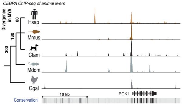
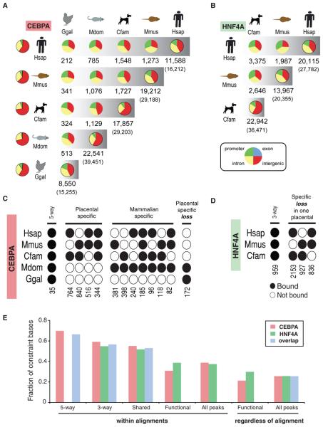
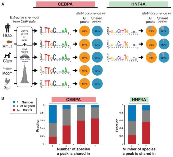
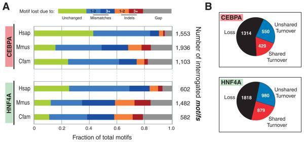
Transcription factors (TFs) direct gene expression by binding to DNA regulatory regions. To explore the evolution of gene regulation, we used chromatin immunoprecipitation with high-throughput sequencing (ChIP-seq) to determine experimentally the genome-wide occupancy of two TFs, CCAAT/enhancer-binding protein alpha and hepatocyte nuclear factor 4 alpha, in the livers of five vertebrates. Although each TF displays highly conserved DNA binding preferences, most binding is species-specific, and aligned binding events present in all five species are rare. Regions near genes with expression levels that are dependent on a TF are often bound by the TF in multiple species yet show no enhanced DNA sequence constraint. Binding divergence between species can be largely explained by sequence changes to the bound motifs. Among the binding events lost in one lineage, only half are recovered by another binding event within 10 kilobases. Our results reveal large interspecies differences in transcriptional regulation and provide insight into regulatory evolution.


Figures

Figure 1
Figure 1
CEBPA binding in vivo in livers isolated from five vertebrate species cross-mapped to the human PCK1 gene locus. A rare ultraconserved binding event is shown surrounded by species-specific and partially-shared binding events. On the left is the evolutionary tree of the five study species (Hsap=Homo sapiens; Mmus=Mus musculus; Cfam=Canus familiaris; Mdom=Monodelphis domesticus; Ggal=Gallus gallus), with their approximate evolutionary distance in millions of years (MY). The bottom track shows evolutionary conservation measured across 44 vertebrate species, and darker shading represents slower evolution.
Figure 2
Figure 2
Conservation and divergence of transcription factor binding. (A) For CEBPA and (B) HNF4A, the pair-wise distribution and numbers of binding events are shown as a pie chart distributed into: intergenic (red), intronic (yellow), exonic (blue), and promoter [TSS +/− 3kb] (green) regions. The left-most column contains the distributions of the bulk genomes. The right-most pie chart represents all binding events in each species with the total number of alignable peaks above the total peaks (in parentheses). (C,D) Multi-species CEBPA and HNF4A binding event analysis where black circles indicate binding in a given species. For instance, there are 764 regions bound by CEBPA only in dog and human (see also Figure S6, S7, S17, and Tables S2, S6). (E) The DNA sequence constraint beneath binding events was measured by average Genomic Evolutionary Rate Profiling (19) scores for peaks found: in all 5 species (5-way) among all the placental mammals (3-way), bound in any two species (Shared), within 10 kb of the TSS of functional targets (Functional), and all peaks.
Figure 3
Figure 3
DNA binding specificities of CEBPA and HNF4A are highly conserved during vertebrate evolution. (A) The known sequence motifs were identified de novo in each species interrogated (Methods), and found within almost all binding events (see Figure S12). (B) Multiple aligned motif occurrences are highly associated with binding events shared among three or more species. Peaks are categorized by the number of species they are shared in and the fraction of peaks with 0 (blue), 1 (grey), and 2 or more (red) aligned motifs are shown.
Figure 4
Figure 4
Lineage-specific loss and turnover of transcription factor binding events. (A) The unbound regions in each placental mammal that align to regions showing TF binding in the other two placental mammals were collected, and the mechanisms by which the underlying motifs were disrupted were summarized. (B) Turnovers occurred near lineage-specific lost binding events approximately half the time; shared turnovers represent cases where a cluster of binding events likely occurred in a common ancestor (see text, Figure S16).

Comment in

References

    1. Wray GA. Nat Rev Genet. 2007;8:206–16. - PubMed
    1. Lenhard B, Sandelin A, Mendoza L, Engström P, et al. J Biol. 2003;2:13. - PMC - PubMed
    1. Boffelli D, McAuliffe J, Ovcharenko D, Lewis KD, et al. Science. 2003;299:1391–4. - PubMed
    1. Borneman AR, Gianoulis TA, Zhang ZD, Yu H, et al. Science. 2007;317:815–9. - PubMed
    1. Dermitzakis ET, Clark AG. Mol Biol Evol. 2002;19:1114–21. - PubMed

Publication types

MeSH terms